Friday Board Review

Board Review by Dr. Edward Guo (Edited by Dr. Parikh)

A 30 year old male presents with a displaced right ankle bimalleolar fracture. He is undergoing procedural sedation in the emergency department using midazolam and fentanyl for fracture-dislocation reduction. During the procedure, he becomes apneic and hypoxic. The hypoxia improves with bag valve ventilation, but he becomes progressively more difficult to ventilate. There is absence of chest rise despite increasing positive pressure. What is the likely cause of this patient’s presentation?

A: Laryngospasm

B: Musculoskeletal stiffness

B: Opioid induced hypoventilation

C: Pneumothorax

Answer: Musculoskeletal stiffness

This patient is likely experiencing Rigid Chest Syndrome, a rare but potentially fatal side effect of synthetic opioids causing skeletal muscle rigidity. The exact mechanism is unknown but is related to the dose and administration. It is commonly seen at high doses (> 3 mcg/kg of fentanyl) and with rapid IV push but has been reported with low doses as well. Treatment includes use of propofol for muscle relaxation or naloxone for reversal of opioid agonism. Neuromuscular paralysis and intubation may be required in refractory cases.

Laryngospasm is a known adverse reaction of ketamine administration which usually responds to first-line maneuvers such as jaw thrust or bag valve ventilation. Hypoventilation is a common side effect of opioids but should not cause chest wall rigidity. While uncommon, a pneumothorax may be caused by excessive positive pressure, but at least unilateral chest rise should be visualized with ventilation.

References:

Myers JG, Sutherland J. Procedural Sedation and Analgesia in Adults. In: Tintinalli JE, Ma O, Yealy DM, Meckler GD, Stapczynski J, Cline DM, Thomas SH. eds. Tintinalli’s Emergency Medicine: A Comprehensive Study Guide, 9e. McGraw-Hill Education; 2020.

Çoruh  B, Tonelli  MR, Park  DR: Fentanyl-induced chest wall rigidity case report. Chest 143: 1145, 2013. [PubMed: 23546488.

Patel, Nishika. “Wooden Chest Syndrome.” CriticalCareNow, 5 Aug. 2021, criticalcarenow.com/wooden-chest-syndrome/. Accessed 22 Mar. 2024.

Friday Board Review

Board Review by Dr. Alex Hilbmann (Edited by Dr. Parikh)

A 66 year old female reports to the emergency department with right arm pain after slipping on ice and trying to catch herself while falling forward. The patient is in incredible pain and has an obvious deformity of the right forearm upon presentation. An x-ray of the right forearm is shown below. What injury does the patient have?

A. Colles Fracture

B. Monteggia Fracture

C. Galeazzi Fracture

D. Smith Fracture

Answer is C. The x-ray shows a distal third radial fracture with disruption of the distal radioulnar joint space, which is a Galeazzi fracture. A Colles Fracture (A) is a distal radius fracture with dorsal displacement whereas a Smith Fracture (D) is a distal radius fracture with volar displacement. Both fractures frequently do not involve significant disruption of the radioulnar joint. A Monteggia fracture (B) is a fracture of the proximal ulna which results in radial head dislocation. For a Galeazzi fracture, the anterior osseous nerve (a branch of the median nerve) is often affected and function should be assessed by asking the patient to perform an “Okay” sign with first and second digit of affected arm.  Orthopedic Surgery consultation is necessary in adults as treatment is an open reduction and internal fixation (ORIF). If open fracture is present Cefazolin should be administered and if the wound is > 10 cm, appears contaminated, or involves seawater/freshwater/farming equipment Gentamicin should also be administered.

Picture from:

https://www.orthobullets.com/trauma/1029/galeazzi-fractures

Resources:

Atesok KI, Jupiter JB, Weiss AP. Galeazzi fracture. J Am Acad Orthop Surg. 2011 Oct;19(10):623-33. doi: 10.5435/00124635-201110000-00006. PMID: 21980027.

Garg R, Mudgal C. Galeazzi Injuries. Hand Clin. 2020 Nov;36(4):455-462. doi: 10.1016/j.hcl.2020.07.006. PMID: 33040957.

Mills, Trevor J.. “Forearm Fractures.” (2013). https://www.semanticscholar.org/paper/Forearm-Fractures-Mills/dead0398468fc50a88349251a8cb8a49b88f838f

Friday Board Review

Board Review by Dr. Guo (Edited by Dr. Parikh)

A 28 year old male presents for finger pain. He works in construction and accidentally poked his right index finger with a stray nail a few days ago. Since then, the palmar aspect of his fingertip has become progressively more swollen and painful. Vital signs are within normal limits. On exam, the right upper extremity is neurovascularly intact with full range of motion. There is erythema, fluctuance, and severe pain to palpation over the distal pulp of his second digit. There is no pain to palpation proximally along the finger. What organism is the most common cause of this patient’s diagnosis?

A: Methicillin-resistant S. aureus (MRSA)

B: Methicillin-sensitive S. aureus (MSSA)

C: Pseudomonas aeruginosa

D: Streptococcus pyogenes

Answer: Methicillin-resistant S. aureus (MRSA)

This patient is presenting with a felon, a subcutaneous pyogenic infection of the distal finger or thumb. The infection typically results from a minor puncture wound which later becomes an abscess confined to the small compartments of the finger pad. Treatment commonly involves incision and drainage in addition to oral antibiotics. Thus, it is extremely important that the antibiotics appropriately cover MRSA as improperly treated felons may worsen to cause flexor tenosynovitis or osteomyelitis. MSSA and Streptococcus pyogenes are other common causes of felons but not as common as MRSA. Pseudomonas is not a common cause of felons.

References:

Wilson  PC, Rinker  B: The incidence of methicillin-resistant Staphylococcus aureus in community-acquired hand infections. Ann Plast Surg 62: 513, 2009. [PubMed: 19387151]  
Germann CA. Nontraumatic Disorders of the Hand. In: Tintinalli JE, Ma O, Yealy DM, Meckler GD, Stapczynski J, Cline DM, Thomas SH. eds. Tintinalli’s Emergency Medicine: A Comprehensive Study Guide, 9e. McGraw-Hill Education; 2020.

Friday Board Review

Board Review by Dr. Edward Guo (Edited by Dr. Parikh)

A 66 year old male with a past medical history of type 2 diabetes presents to the emergency department for head injury. He was the restrained driver when he swerved his car to avoid a child that ran into the street to retrieve a ball. He was driving approximately 15 mph in his neighborhood and swerved onto grass. He hit his head on the driver side window and complains of a headache. He was able to ambulate out of the vehicle and denies loss of consciousness, vomiting, or use of blood thinners. Vital signs are normal. On exam, he is neurologically intact and has ecchymosis to the left forehead. Which of the following is appropriate justification to obtain head imaging according to the Canadian CT Head Rule?

A: Dangerous mechanism

B: Headache

C: Patient age 

D: Rule does not apply to this patient

Answer: Rule does not apply to this patient

The Canadian CT Head Rule is a widely used clinical decision tool that emergency physicians frequently utilize to screen for significant head injuries. In the external validation trial, it was found to be 100% sensitive in detecting both clinically important brain injuries and injuries that required neurosurgical intervention. This patient does not meet the inclusion criteria for its use. If he did meet the inclusion criteria, his age would be an appropriate justification of head imaging as a high risk criteria. Headache is not part of the decision tool. His injury also does not meet the medium risk dangerous mechanism. 

MDCalc states, “Patients with minimal head injury (i.e., no history of loss of consciousness, amnesia, and confusion) generally do not need a CT scan. For example, patients over 65 years old may not need a CT scan just based on their age if they do not have the history mentioned above. When a patient fails the CCHR, use clinical judgment on whether a CT scan is necessary.”

Canadian CT Head Rule
Inclusion CriteriaExclusion Criteria
Head injury with GCS 13-15 and at least one of the following:Age < 16 years
    Loss of consciousnessUse of blood thinners
    Amnesia to head injury eventSeizure after injury
    Witnessed disorientation

References:

Stiell IG, Wells GA, Vandemheen K, et al. The Canadian CT Head Rule for patients with minor head injury. Lancet. 2001;357(9266):1391-1396. doi:10.1016/s0140-6736(00)04561-x

https://www.mdcalc.com/calc/608/canadian-ct-head-injury-trauma-rule

Friday Board Review

Board Review by Dr. Guo (Edited by Dr. Parikh)

A 30 year old obese female presents to the emergency department for a persistent headache. You have a high suspicion for idiopathic intracranial hypertension and perform a lumbar puncture. The opening pressure is 28 cm H2O. Approximately how much volume of CSF should be removed to reach a target CSF pressure of 20 cm H2O prior to removal of the spinal needle?

A: 4 mL

B: 8 mL

C: 16 mL

D: 20 mL

Answer: 8 mL

Part of the diagnostic criteria for idiopathic intracranial hypertension in adults includes an elevated opening pressure > 25 cm H2O on lumbar puncture. The feared complication is permanent vision loss from papilledema. Treatment includes removal of CSF which can also provide relief of headache symptoms. As a general rule, removal of 1 mL of CSF will lower the CSF pressure by about 1 mL H2O. It is recommended to remove the desired amount of CSF and then re-measure the CSF pressure prior to removal of the spinal needle. Excess removal of CSF can result in intracranial hypotension and a low pressure headache.

References:

Fiorito-Torres  F, Rayhill  M, Perloff  M: Idiopathic intracerebral hypertension (IIH)/pseudotumor: removing less CSF is best (I9-1.006). Neurology 82 (10 Suppl): I9–1.006, 2014.

Koyfman A, Long B. Headache. In: Tintinalli JE, Ma O, Yealy DM, Meckler GD, Stapczynski J, Cline DM, Thomas SH. eds. Tintinalli’s Emergency Medicine: A Comprehensive Study Guide, 9e. McGraw-Hill Education; 2020.

Thurtell, Matthew & Bruce, Beau & Newman, Nancy & Biousse, Valérie. (2010). An Update on Idiopathic Intracranial Hypertension. Reviews in neurological diseases. 7. e56-68. 10.3909/rind0256.

Friday Board Review

Board Review by Dr. Guo (Edited by Dr. Parikh)

A 40 year old male with a past medical history of polysubstance use, epilepsy, and housing insecurity presents to the emergency department for drug intoxication. His ED course is uncomplicated, and he is deemed ready for discharge 3 hours after his initial presentation. Upon handing him his discharge paperwork, he appears to trip and fall to the ground. He then demonstrates diffuse shaking of his entire body with his eyes tightly shut, not responding to external stimuli. The episode lasts approximately 2 minutes, and he afterward remains unresponsive to stimuli. Vital signs and point-of-care glucose are within normal limits. Physical exam shows no obvious injuries. Which of the following laboratory tests is most helpful in determining if the patient had an epileptic seizure?

A: Creatine kinase

B: Lactic acid

C: Potassium

D: White blood cell count

Answer: Lactic acid

Distinguishing between true epileptic seizures, psychogenic non-epileptic seizures (PNES), and convulsions following a syncopal episode can be difficult. PNES is psychogenic in nature and has highly variable features which may include forceful closing of the eyelids, side-to-side movements, or shrieking. An elevated serum lactic acid concentration obtained shortly after the event has been shown to help differentiate true epileptic seizures from PNES or convulsions following syncope. Creatine kinase levels typically do not rise early after a seizure and are furthermore not specific in the setting of falls or trauma. Potassium levels are not expected to be elevated following an uncomplicated seizure. An elevated white blood cell count may be due to several non-specific reasons including infection, trauma, steroid use, or stress response. Definitive diagnosis is determined using electroencephalography (EEG).

References:

Kornegay J. Seizures and Status Epilepticus in Adults. In: Tintinalli JE, Ma O, Yealy DM, Meckler GD, Stapczynski J, Cline DM, Thomas SH. eds. Tintinalli’s Emergency Medicine: A Comprehensive Study Guide, 9e. McGraw-Hill Education; 2020.

Patel J, Tran QK, Martinez S, Wright H, Pourmand A. Utility of serum lactate on differential diagnosis of seizure-like activity: A systematic review and meta-analysis. Seizure. 2022;102:134-142. doi:10.1016/j.seizure.2022.10.007

Friday Board Review

Board Review by Dr. Edward Guo (Edited by Dr. Parikh)

A 30 year old male with a history of cardiac arrest with ischemic encephalopathy status post tracheostomy and gastrostomy placement presents from a long-term care facility for a feeding tube problem. His nurse was bathing and performing dressing changes when the patient’s gastrostomy tube fell out. He has otherwise had no fever or vomiting, and his last bowel movement was earlier today. Vital signs are within normal limits. On exam, he appears comfortable. Patient is non-verbal and does not follow commands. There is a patent gastrostomy stoma in his left upper quadrant with no surrounding erythema or drainage. Old charts state that general surgery created the gastrostomy 6 weeks ago with a 16-french tube. Which of the following is the most appropriate initial management?

A: Consult General Surgery for gastrostomy tube replacement

B: CT abdomen and pelvis

C: insert a 14-french gastrostomy tube

D: insert a 16-french gastrostomy tube

Answer: Insert a 16-french gastrostomy tube

Artificial stomas are at risk for premature closure if the tube has been accidentally removed. Closure may begin quickly (within hours) depending on how mature the tract is. It is important for the emergency physician to be knowledgeable of the maturity of different surgical stomas and when consultation is necessary. In general, gastrostomy tracts mature after 2 to 3 weeks and then afterward can be replaced in the emergency department. Using the previous size tube is preferred to prevent leakage around the tract with a smaller diameter tube. CT of the abdomen and pelvis is unlikely to change management given the patient is asymptomatic. If the gastrostomy tract is immature or a 16-french tube is difficult to insert, then it would be indicated to consult general surgery for replacement. Do not attempt to push through resistance due to the risk of creating a false tract. In that case, attempting to insert a smaller size tube is advised to keep the original tract patent.

Type of Surgical StomaTime to Mature
Tracheostomy7 to 10 days
Gastrostomy2 to 3 weeks
Cystostomy (suprapubic)4 to 6 weeks (little evidence, varies based on provider)

References:

Witting MD. Gastrointestinal Procedures and Devices. In: Tintinalli JE, Ma O, Yealy DM, Meckler GD, Stapczynski J, Cline DM, Thomas SH. eds. Tintinalli’s Emergency Medicine: A Comprehensive Study Guide, 9e. McGraw-Hill Education; 2020.
Buscaglia  JM: Common issues in PEG tubes—what every fellow should know. Gastrointest Endosc 64: 970, 2006. [PubMed: 17140906]

Friday Board Review

Board Review by Dr. Guo (Edited by Dr. Parikh)

A 40 year old female with a history of kidney stones with a left ureteral stent placed 2 years ago presents for urinary pain associated with increased urge and frequency. She denies fever or flank pain. It does not feel similar to her previous kidney stones. Vital signs are within normal limits. On exam, she is comfortable appearing and has mild suprapubic tenderness to palpation. Point-of-care pregnancy test is negative. Urinalysis results with 3+ leukocyte esterase, 2+ nitrites, and 105 WBCs/HPF. Which of the following is the appropriate management of this patient’s condition?

A: Admit for IV antibiotics

B: Consult urology for stent removal

C: CT of the abdomen and pelvis

D: Discharge with oral antibiotics

Answer: discharge with oral antibiotics

This patient is presenting with findings consistent with acute cystitis, a urinary tract infection localized to the bladder. Urinalysis findings with elevated leukocyte esterase, nitrites, and WBCs with clinical symptoms are supportive of the diagnosis. Treatment of minor urinary tract infections in patients with ureteral stents is oral antibiotics and do not require stent removal. If pyelonephritis or systemic infection with a ureteral stent is suspected, consultation with urology, IV antibiotics, and radiographic imaging to determine the location of the stent is warranted. 

References:

Askew KL. Urinary Tract Infections and Hematuria. In: Tintinalli JE, Ma O, Yealy DM, Meckler GD, Stapczynski J, Cline DM, Thomas SH. eds. Tintinalli’s Emergency Medicine: A Comprehensive Study Guide, 9e. McGraw Hill; 2020.

Josephson EB, Azan B. Complications of Urologic Procedures and Devices. In: Tintinalli JE, Ma O, Yealy DM, Meckler GD, Stapczynski J, Cline DM, Thomas SH. eds. Tintinalli’s Emergency Medicine: A Comprehensive Study Guide, 9e. McGraw Hill; 2020.

Friday Board Review

Board Review by Dr. Hilbmann (Edited by Dr. Parikh)

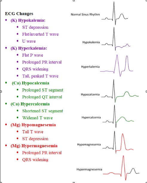
A 22 year old female G1P0 with no pmhx who presents to the Emergency Department with nausea and vomiting. The patient is 11 weeks pregnant and has experienced nausea and occasional vomiting throughout her pregnancy but for the past three days she has been progressively worse and has not been able to keep any food or liquids down without vomiting. You are given her ECG, what is a finding you are looking for due to her recent presentation?


A. Hyperacute T waves
B. Shortened PR Interval
C. Prolonged QT interval
D. ST-Elevation in aVR

Answer: C.
Due to this patient’s increased vomiting, most likely due to her pregnancy, it is possible that she may be experiencing electrolyte imbalances secondary to the loss of gastrointestinal contents. Most commonly hypokalemia, hypocalcemia, and hypomagnesemia can result from continuous vomiting. In addition to blood tests, which take time to result, the levels of these three electrolytes can be evaluated with ECG as well. Physicians should evaluate for increased p waves, prolonged PR interval, ST depression, u waves, and a prolonged QT interview.

References:
Mitchell SJ, Cox P. ECG changes in hyperemesis gravidarum. BMJ Case Rep. 2017;2017(bcr2016217158) doi: 10.1136/bcr-2016-217158.
Popa SL, Barsan M, Caziuc A, Pop C, Muresan L, Popa LC, Perju-Dumbrava L. Life-threatening complications of hyperemesis gravidarum. Exp Ther Med. 2021 Jun;21(6):642. doi: 10.3892/etm.2021.10074. Epub 2021 Apr 16. PMID: 33968173; PMCID: PMC8097228.
Image per @medicalce via Twitter

Friday Board Review

Board Review by Alex Hilbmann

Vital signs:

T: 37⁰C HR: 71 bpm O2 Sat: 100% BP: 112/92

A 29 year old female reports to the emergency department for abnormal scant vaginal bleeding. Initially, she believed that the bleeding was an early menstrual period but it has now persisted longer than her usual menses with less volume. She denies any other complaints, including pelvic pain, fevers, or vaginal discharge. Patient has attended OB/GYN appointments yearly and denies any previous history of sexually transmitted infections or abnormal pap smears. Pelvic exam reveals scant bleeding from the cervical os with no adnexal or cervical tenderness. No masses are appreciated upon palpation of bilateral adnexa. Transvaginal ultrasound reveals no intrauterine pregnancy or adnexal abnormalities. Point of care urine pregnancy test is positive. Quantitative beta-hcg results at 542 miU/mL. Vital Signs listed above. What is the next best step in management of this patient?

  1. Consultation to OB/GYN for concern of ectopic pregnancy
  2. Official transvaginal ultrasound read by Radiology
  3. Administration of methotrexate and discharge home
  4. Discharge home with 2 day OB/GYN follow up

Answer: D.

Our patient is currently experiencing scant vaginal bleeding with no findings on transvaginal ultrasound, a positive pregnancy test, and a beta hcg below 1500 miU/mL.  1500 miU/mL is what is known as the “discriminatory zone” for transvaginal ultrasound (6,000 miU/mL for transabdominal). This discriminatory zone is a level of beta-hcg which an intrauterine pregnancy (IUP) would be expected to be seen on ultrasound. When beta-hcg is above the discriminatory zone, and therefore an IUP should be visible on ultrasound, the absence of an IUP on ultrasound suggests ectopic pregnancy until proven otherwise. With this patient however, we are below the discriminatory zone and therefore the absence of IUP (with no other findings suggesting ectopic pregnancy) does not make ectopic pregnancy a more likely diagnosis than pregnancy with implantation bleeding at this time. The beta hcg of an IUP should double by two days, whereas an ectopic pregnancy would decrease less than twofold. This can be assessed by the patient’s OB/GYN at her follow up, and given the patient’s stable condition she can be discharged home.

Tintinalli, J., Ma, O., Yealy, D., Meckler, G., Cline, D., Thomas, S. and Stapczynski, J., 2020. Tintinalli’s emergency medicine. 9th ed. [New York]: McGraw-Hill Education, pp.615-620.

Image:

Fadial, T., 2018. Differential Diagnosis of Ultrasound in Ectopic Pregnancy. [online] Differential Diagnosis of. Available at: <https://ddxof.com/ultrasound-in-ectopic-pregnancy/?sf_action=get_data&sf_data=all&_sf_s=ectopic> [Accessed 2 October 2022].