Friday Board Review

Internal Medicine Board Review with Dr. Edward Guo

A 30 year old female with a history of type 1 diabetes and past hospitalizations for diabetic ketoacidosis presents via EMS for altered mental status. History is limited as patient is altered and not answering questions appropriately. Vitals include Temp 100.4F, HR 116, BP 102/70, RR 30, SpO2 98% on room air. Exam shows an ill-appearing female with Kussmaul respirations and a non-focal neurologic exam. Labs are notable for 20K WBCs and serum glucose of 400. A lumbar puncture is performed to assess for meningitis. For this patient, which of the following CSF glucose values is within normal limits?

A: 60 mg/dL

B: 100 mg/dL

C: 260 mg/dL

D: 400 mg/dL

Answer: 260 mg/dL

This patient is presenting with signs and symptoms of diabetic ketoacidosis. While it is critical for the emergency physician to treat the hyperglycemia with volume resuscitation and insulin, it is also paramount to investigate for underlying causes such as infection. The glucose level in CSF is proportional to serum glucose values and should correspond to approximately 60-70% of serum glucose values. Thus, a CSF glucose value of 60 or 100 mg/dL in this patient is lower than expected and concerning for bacterial CNS infection. Higher than expected CSF glucose levels are non-specific and generally do not exceed 300 mg/dL.

References:

Lillian A. Mundt; Kristy Shanahan (2010). Graff’s Textbook of Routine Urinalysis and Body Fluids. Lippincott Williams & Wilkins. p. 237. ISBN 978-1582558752.

Seehusen DA, Reeves MM, Fomin DA (September 2003). “Cerebrospinal fluid analysis”Am Fam Physician68 (6): 1103–8. PMID 14524396

Friday Board Review

Infectious Disease Board Review with Dr. Edward Guo

A 74 year old male with a past medical history of advanced dementia and type 2 diabetes presents via EMS from his long term advanced care facility for cough and shortness of breath. Patient is AOx1 and intermittently follows commands at baseline. EMS reports that the facility nurse noticed that he was hypoxic and had a “nasty cough.” Vitals include Temp 100.6F, HR 110, BP 126/80, RR 22, SpO2 89% on room air. Exam shows a chronically ill, pale appearing older male in mild respiratory distress with a productive cough. Lung sounds are notable for crackles in the lower right lung. A chest x-ray demonstrates focal consolidations of the right middle and right lower lobes with a moderate sized pleural effusion above the right hemidiaphragm. Which of the following laboratory values is NOT part of the diagnostic criteria for an empyema?

A: pleural gram stain of culture

B: pleural LDH

C: pleural pH

D: pleural protein

Answer: pleural protein

This patient is presenting with pneumonia demonstrated by imaging results consistent with the clinical findings of fever, cough, and hypoxia. Pneumonia is the most common cause of an empyema which has specific diagnostic criteria distinct from the Light Criteria for pleural effusions. Approximately 40% of cases have negative cultures. Treatment is drainage and broad spectrum antibiotics with anaerobic coverage.

Diagnostic Criteria for EmpyemaLight Criteria for Exudative Pleural Effusion (requires 1 of the following)
Aspiration of grossly purulent fluid plus one of the following:Pleural protein/serum protein > 0.5
     Positive gram stain or culturePleural LDH/serum LDH > 0.6
     Pleural fluid glucose < 40Pleural LDH > 2/3 upper limit of normal serum LDH
     Pleural pH < 7.2 
     Pleural LDH > 1000 

References:
Mace SE, Anderson E. Lung Empyema and Abscess. In: Tintinalli JE, Ma O, Yealy DM, Meckler GD, Stapczynski J, Cline DM, Thomas SH. eds. Tintinalli’s Emergency Medicine: A Comprehensive Study Guide, 9e. McGraw Hill; 2020.

Birkenkamp  K, O’Horo  JC, Kashyap  R,  et al: Empyema management: a cohort study evaluating antimicrobial therapy. J Infect 72: 537, 2016.

Friday Board Review

From the Archives: Pediatrics Board Review with Dr. Christine Collins

An 8 month old male born at 36 weeks without any complications with no medical problems who presents with wheezing, increased work of breathing, rhinorrhea and cough for the past 2 days. On exam, he has a low grade temperature, wheezing in all lung fields, subcostal retractions and nasal flaring. HR is 156 bpm, RR 70, Oxygen saturation is 90% on room air. Mother says other siblings in the house have had a cold the past few days. What is the next step in the management of this patient?

a. Administer IV dexamethasone

b. Administer broad spectrum IV antibiotics

c. Admit with supportive measures

d. Administer inhaled corticosteroids

Answer: Admit with supportive measures

This patient is presenting with acute signs and symptoms of bronchiolitis which include rhinorrhea, cough, wheezes, cough, crackles, use of accessory muscles, and nasal flaring. Babies born prematurely are at increased risk for severe bronchiolitis. Clinically, bronchiolitis occurs primarily <2 years of age, with a peak presentation between 6 and 12 months. 

Treatment for bronchiolitis includes supportive care measures: nasal suctioning and saline drops, oxygen, isotonic fluids, and ventilatory support if needed. Consider hospitalization if persistent increased work of breathing, inability to maintain hydration/feeding, or hypoxia. Beta agonists can be trialed if the patient has a family history suggestive of asthma or atopy. Corticosteroids are not recommended for routine use.

References:

Bjoernsen, L. P., & Ebinger, A. (2016). Chapter 124 Wheezing in Infants and Children In Tintialli’s Emergency Medicine: A Comprehensive Study Guide (8th ed). New York, NY: McGraw-Hill Education.

Cade A, Brownlee KG, Conway SP, et al. Randomised placebo controlled trial of nebulised corticosteroids in acute respiratory syncytial viral bronchiolitis. Arch Dis Child 2000; 82:126

Friday Board Review

Pharmacology Board Review with Dr. Edward Guo

A 30 year old non-pregnant female presents after a cat bite to her left hand. Vitals are within normal limits. The extremity is neurovascularly intact and shows two subcentimeter punctate lesions on the palmar surface of her hand. She reports an anaphylactic reaction to penicillin. Which of the following antibiotics is appropriate prophylactic treatment for this patient?

A: amoxicillin-clavulanate

B: clindamycin

C: cephalexin

D: doxycycline

Answer: doxycycline

Prophylactic antibiotic treatment is common practice of mammalian bites in the ED. Antibiotics should be tailored for coverage of streptococcal and staphylococcal bacteria as well as anaerobes and Pasteurella multocida. Amoxicillin-clavulanate is typically the antibiotic of choice but is contraindicated in patients with an anaphylactic allergy to penicillin. Clindamycin or cephalexin monotherapy do not reliably cover Pasteurella species. Doxycycline or dual therapy with clindamycin plus TMP-SMX or a fluoroquinolone are reasonable alternatives for penicillin-allergic patients.

References:

Quinn J (2020). Puncture wounds and bites. Tintinalli J.E., & Ma O, & Yealy D.M., & Meckler G.D., & Stapczynski J, & Cline D.M., & Thomas S.H.(Eds.), Tintinalli’s Emergency Medicine: A Comprehensive Study Guide, 9e. McGraw Hill.

Presutti R. J. (2001). Prevention and treatment of dog bites. American family physician63(8), 1567–1572.

Friday Board Review

Cardiology Board Review by Dr. Alex Hilbmann

You are asked to see a patient with a known history of Left Ventricular Assist Device (LVAD) due to end stage heart failure who presents after becoming more confused in the Emergency Department waiting room. Per family the patient was originally presenting due to feeling lightheaded/fatigued recently. The patient does not have a pulse and the automatic/manual blood pressure cuff does not determine a blood pressure. Patient cannot follow commands but localizes to pain in all four extremities. Capillary refill < 2 seconds. What should be done next?

A. Begin chest compressions

B. Auscultate heart

C. Point of care echocardiogram

D. Portable CXR

Answer: Auscultate heart

The first thing that should be done for this patient is auscultate his heart to note if an audible “whirr” is present. If it is present, this would indicate that his LVAD is in fact operating. Patients who have an LVAD do not have a palpable pulse/blood pressure therefore in this population it does not indicate that their LVAD is currently not working/they are not perfusing. An ultrasound with doppler may be used to obtain a blood pressure reading. Chest compressions should be withheld until absolutely necessary given the chance to potentially dislodge the LVAD causing dysfunction or left ventricular rupture. The chance of chest compressions causing damage to LVAD can be minimized by paying attention to positioning of compressions and avoiding unnecessarily deep compressions. It would be reasonable to begin compressions if low perfusion state suggested by exam (mental status change, skin mottling, capillary refill >2 seconds). Auscultation of the heart would be the most timely and effective method in recognizing functionality of patient’s LVAD. Given patient’s exam concerning for altered mental status without other findings for low perfusion, CPR can briefly be held until further evaluation determines concern for hypoperfusion.

References:

Guglin, Maya (2018) “Approach to Unresponsive Patient with LVAD,” The VAD Journal: Vol. 4, Article 2.
DOI: https://doi.org/10.13023/VAD.2018.02
Available at: https://uknowledge.uky.edu/vad/vol4/iss1/2

Tintinalli, J., Ma, O., Yealy, D., Meckler, G., Cline, D., Thomas, S. and Stapczynski, J., 2020. Tintinalli’s emergency medicine. 9th ed. [New York]: McGraw-Hill Education, pp.382

Yuzefpolskaya M, Uriel N, Flannery M, Yip N, Mody K, Cagliostro B, Takayama H, Naka Y, Jorde UP, Goswami S, Colombo PC. Advanced cardiovascular life support algorithm for the management of the hospitalized unresponsive patient on continuous flow left ventricular assist device support outside the intensive care unit. Eur Heart J Acute Cardiovasc Care. 2016 Dec;5(8):522-526. doi: 10.1177/2048872615574107. Epub 2015 Mar 4. PMID: 25740223.

Friday Board Review

Board Review with Dr. Edward Guo

A 30 year old male with a history of active IVDU and previous MRSA endocarditis is presenting with tooth pain that has been worsening over several days. He denies fever, chest pain, or shortness of breath. Vital signs are: Temp 99.0F, HR 86, BP 148/76, RR 16, SpO2 98% RA. Exam shows track marks in the antecubital fossas bilaterally and no appreciable cardiac murmur. He has poor dentition overall with an appreciable area of fluctuance above the gums of tooth #4. Which of the following is appropriate management for this patient?

A: administer IV vancomycin followed by ED incision and drainage then discharge

B: consult oral maxillofacial surgery for drainage

C: draw blood cultures and admit for IV antibiotics

D: perform ED incision and drainage and discharge with clindamycin

Answer: administer IV vancomycin followed by ED incision and drainage then discharge

Patients with a history of prosthetic heart valves or infective endocarditis among other cardiac conditions are considered high-risk for developing endocarditis with dental procedures and surgical procedures on infected skin. In this patient, incision and drainage of the periapical abscess should be performed 30 to 60 minutes after receiving a dose of antibiotics with coverage against MRSA. OMFS does not need to be consulted for abscess drainage. There are no systemic symptoms such as fever at this time to suggest bacteremia for admission. 

Table copied from Tintinalli’s

References:

Brenner D, & Marco C.A., & Rothman R.E. (2020). Endocarditis. Tintinalli J.E., & Ma O, & Yealy D.M., & Meckler G.D., & Stapczynski J, & Cline D.M., & Thomas S.H.(Eds.), Tintinalli’s Emergency Medicine: A Comprehensive Study Guide, 9e. McGraw Hill.

Friday Board Review

From the EMDaily Archives: Board Review by Dr. Courtney Martin

A 50 year old male with past medical history of hypertension, type 2 diabetes, and coronary artery disease with multiple prior stents presenting with shortness of breath and fatigue. Vitals demonstrate BP 70/50, HR 79 bpm, RR 26, O2 sat 88% on room air, Temp 36C. On exam, the patient has bilateral rales and 2+ pitting edema. Heart sounds are muffled. Distal pulses are weak, and extremities are cool to the touch. Bedside echocardiogram demonstrates significantly decreased cardiac output with minimal ventricular wall motion. EKG demonstrates ST elevations in anterior leads. What is the most appropriate vasopressor to initiate at this time?

A: dobutamine

B: dopamine

C: epinephrine

D: norepinephrine

E: phenylephrine

Answer: norepinephrine

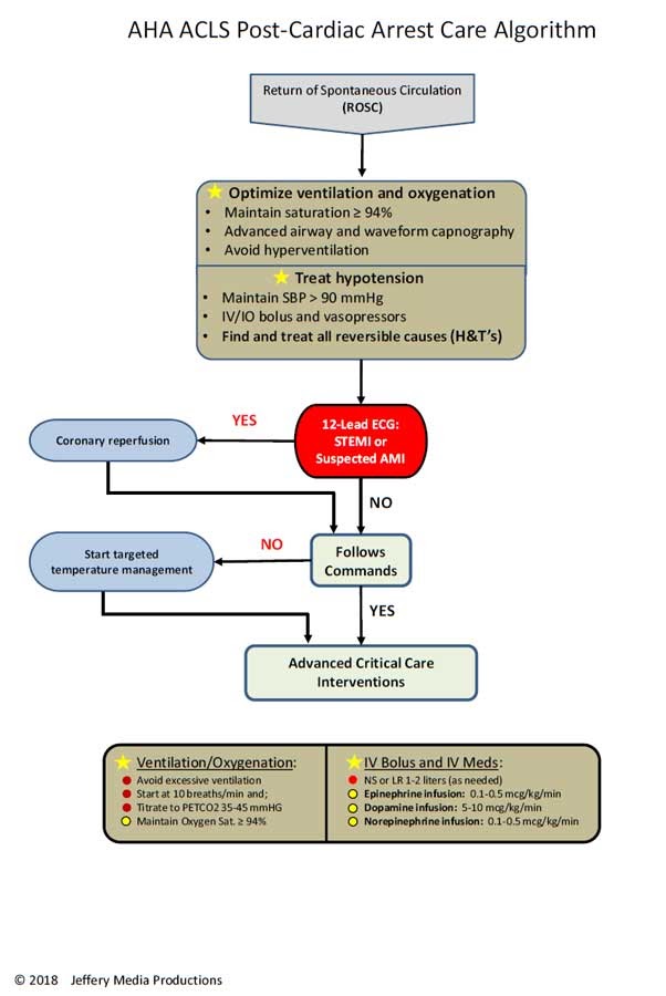
This patient is demonstrating signs of cardiogenic shock in the setting of acute myocardial infarction. The most common cause of cardiogenic shock is acute ischemia. Other causes include valvular malfunctions, ventricular septal defect, ventricular free-wall rupture, right ventricular infarction, myocarditis, septic shock, arrhythmia, toxicity. The initial step in this case is correction of acute ischemia by activating the cath lab or thrombolytics depending on your facility. In regards to vasopressors, studies have demonstrated norepinephrine is most beneficial in patients with profound hypotension. Dopamine was traditionally used, however, it may increase heart rate and worsen cardiac demand. Epinephrine should be avoided if possible due to increased vasoconstriction with ischemia and arrhythmias. Dobutamine has inotropic effects that are beneficial for cardiac output but does not improve systemic vascular resistance. The combination of norepinephrine and dobutamine may be considered as well. 

References:

Hochman JS, Buller CE, Sleeper LA, et al. Cardiogenic shock complicating acute myocardial infarction–etiologies, management and outcome: a report from the SHOCK Trial Registry. SHould we emergently revascularize Occluded Coronaries for cardiogenic shocK?. J Am Coll Cardiol. 2000;36(3 Suppl A):1063-1070. doi:10.1016/s0735-1097(00)00879-2

Friday Board Review

Board Review with Dr. Edward Guo

A 70 year old male with a past medical history coronary artery disease, heart failure with reduced ejection fraction, and severe aortic stenosis presents via EMS for shortness of breath. History is limited due to acute respiratory distress while patient is on CPAP. Vital signs are: BP 88/60, HR 120, T 36.7, RR 30, SpO2 90% on PEEP 8 and FiO2 100%. On exam, he is in severe respiratory distress with accessory muscle usage and speaks in 2 word phrases. There is a prominent systolic ejection murmur over the right second intercostal space. Rales are heard at the lung bases bilaterally, and there is 4+ pitting edema of the lower extremities. EKG shows sinus tachycardia. Which of the following is the preferred resuscitation strategy to optimize hemodynamics prior to intubation? 

A: bolus 1 liter isotonic fluids

B: epinephrine infusion 

C: norepinephrine infusion

D: phenylephrine infusion

E: push dose epinephrine prior to induction

Answer: phenylephrine infusion

This patient is presenting in acute hypoxic respiratory failure likely secondary to pulmonary edema related to acute on chronic heart failure. Patients with severe aortic stenosis are preload dependent to maintain coronary and systemic perfusion. Thus, typical management with positive airway pressure and nitrates should be used cautiously. In hypotensive patients with aortic stenosis, phenylephrine is the vasopressor of choice due to its pure alpha-1 agonist effects to increase diastolic blood pressure and coronary perfusion. Reflex bradycardia is also beneficial to allow for more diastolic filling time. Inotropes such as epinephrine are not recommended due to tachycardia and increased myocardial oxygen demand. Norepinephrine is a reasonable alternative but not the preferred agent. Fluid administration is likely to worsen this patient’s hemodynamics and respiratory status by volume overload.

References:

Goertz AW, Lindner KH, Schutz W, Schirmer U, Beyer M, Georgieff M. Influence of phenylephrine bolus administration on left ventricular filling dynamics in patients with coronary artery disease and patients with valvular aortic stenosis. Anesthesiology. 1994;81(1):49-58.

Friday Board Review

Board Review with Dr. Edward Guo

A 64 year old male with a past medical history of diabetes mellitus, coronary artery disease, and congestive heart failure is being evaluated for chest pain. His initial vital signs are within normal limits. His EKG is unchanged from previous showing a narrow-complex sinus rhythm. While he is in the emergency department, he reports feeling palpitations. The cardiac monitor records a monomorphic, wide-complex tachycardia at a rate of 140 beats/minute while his blood pressure is 132/80. What is the first-line medication for treatment of this patient’s condition?

A: adenosine

B: amiodarone

C: epinephrine

D: magnesium sulfate

E: procainamide

Answer: procainamide

This patient with a history of extensive cardiac disease is likely experiencing stable ventricular tachycardia (VT). The PROCAMIO study in 2017 demonstrated that procainamide is likely superior to amiodarone for termination of wide-complex tachycardia and associated with fewer major adverse cardiac events. Adenosine is commonly used in the management of supraventricular tachycardias but is unlikely to terminate a tachycardia of ventricular origin. Epinephrine is used for pulseless VT as part of the ACLS algorithm. Magnesium sulfate is used for polymorphic VT also known as Torsades De Pointes but is not first-line for monomorphic VT.

References:

Ortiz, M., Martín, A., Arribas, F., Coll-Vinent, B., Del Arco, C., Peinado, R., Almendral, J., & PROCAMIO Study Investigators (2017). Randomized comparison of intravenous procainamide vs. intravenous amiodarone for the acute treatment of tolerated wide QRS tachycardia: the PROCAMIO study. European heart journal38(17), 1329–1335. https://doi.org/10.1093/eurheartj/ehw230

Friday Board Review

Board Review with Dr. Alex Hilbmann

A 28 year old female G4P2 at 8 weeks gestation presents to the Emergency Department after vomiting almost four times daily for the past week. She denies any recent fevers, abdominal pain, pelvic pain, or vaginal bleeding. Vital signs include: Temp 37.2 C, HR 105, BP 93/62, SpO2 100%. On exam, she is uncomfortable appearing with dry mucous membranes and intermittently dry heaving into an emesis bag. Blood serum results are pending. Urinalysis reveals 1+ ketones with elevated specific gravity. What is the next best step in management?

A. 0.9% normal saline

B. Prophylactic electrolyte repletion

C. 5% dextrose and 0.9% normal saline

D. Antiemetic and PO challenge

Answer: 5% dextrose and 0.9% normal saline

This pregnant patient is most likely experiencing hyperemesis gravidarum given her presentation of multiple episodes of vomiting, volume depletion, and ketonuria. The treatment for hyperemesis gravidarum includes 5% glucose in IV fluids, anti-emetic drugs, and correction of any electrolyte abnormalities. Nothing should be given by mouth until patient’s nausea is controlled, and although this patient will ultimately benefit from antiemetic administration with the hopes that she will tolerate PO, her signs of volume depletion and ketonuria suggest immediate treatment with 5% dextrose in 0.9% normal saline or lactated ringer solution.

Management of Hyperemesis Gravidarum
First line: pyridoxine (vitamin B6) – pregnancy drug class A
Add on: doxylamine – pregnancy drug class A
Adjuncts: ondansetron, metoclopramide – pregnancy drug class B
IV fluids with dextrose

References:

References: Tintinalli, J., Ma, O., Yealy, D., Meckler, G., Cline, D., Thomas, S. and Stapczynski, J., 2020. Tintinalli’s emergency medicine. 9th ed. [New York]: McGraw-Hill Education, pp.621-622.

Nausea and vomiting of pregnancy. ACOG Practice Bulletin No. 153. American College of Obstetricians and Gynecologists. Obstet Gynecol. 2015; 126(3):e12-24