Wednesday Image Review

What’s the Diagnosis? By Dr. Julie Calabrese

75 y/o M PMHx of ESRD on HD, pulmonary HTN, HLD presents to the ED with 1 week of progressive fatigue and SOB. Pt on 2L NC home O2 but requiring 4L NC in the ED to maintain saturation > 95%. On exam, pt with increased WOB and RR > 20. Lungs are CTA. Cardiac exam shows RRR with mild JVD, abdominal distention and +1 pitting edema B/L. POCUS was performed and is shown below. What is the diagnosis? 

Answer: Right Heart Strain from Pulmonary Hypertension 

  • Signs in POCUS that are indicative of R heart strain:
    • D-sign: septal flattening seen in the parasternal short orientation that is indicative of increased RV pressures 
    • McConnel’s Sign: seen in the apical 4 chamber view. R ventricular free wall akinesis with sparing of the apex (apical hyperkinesis) 
    • Increased RV:LV ratio, typically should be ⅓:⅔ 
    • Decreased TAPSE: measurement of the vertical motion of the tricuspid valve in the apical 4 chamber view (normal > 16 mm)
  • Causes of R- Heart Strain:
    • Pulmonary Embolism
    • Pulmonary hypertension 
    • Biventricular failure
    • R sided heart failure 
    • Valvular dysfunction (Acute TR) 
  • Pulmonary Hypertension:
    • Type 1: primary arterial pulmonary HTN 
    • Type 2: PH due to L heart failure
    • Type 3: PH due to lung disease 
    • Type 4: PH due to chronic thromboembolic disease 
    • Type 5: idiopathic PH 
  • Acute Treatment for PH includes
    • Optimize RV preload- patients typically euvolemic or hypervolemic and do not respond well to rapid shifts in fluid status (usually avoid fluids). If hypovolemia/sepsis consider small 250 ml boluses with frequent reassessments 
    • Improve cardiac output: consider early ionotropes 
    • Reduce RV afterload: avoid hypoxia, acidosis, hypercapnia 
    • Treat arrhythmias: most common is SVT followed by afib/flutter 

Resources: 

https://courses.coreultrasound.com/courses/take/fundamentals/lessons/18316427-right-heart-strain-5minsono

Wednesday Image Review

What’s the Diagnosis? By Dr. Carlos Cevallos

A 39 y.o. woman who is G9P1 and currently 6 weeks pregnant presents to the ED with a chief complaint of vaginal bleeding that began in the morning with associated lower abdominal pain and lightheadedness. Physical exam demonstrates lower abdominal tenderness without peritonitis and a small amount of blood in the posterior vaginal fossa with a closed cervical os. You obtain a serum HCG which is 8,960 and perform a transvaginal ultrasound which demonstrates the following. What’s the diagnosis?

Answer: Ectopic Pregnancy

  • When performing a pelvic US in the ED, the focused question is: “Is there an intrauterine pregnancy (IUP) or not?”
  • To diagnose an IUP, one must visualize a gestational sac AND either a yolk sac or fetal pole within the uterus.
  • In this patient, no gestational sac nor yolk sac are visualized within the uterus.
  • The left adnexa demonstrates a tubal ring concerning for an ectopic pregnancy. OBGYN was consulted who took the patient Level 1 to the OR where the ectopic pregnancy was confirmed and removed along with a left salpingectomy.

Resources:

Heaton, Heather. “Chapter 98: Ectopic Pregnancy and Emergencies in the First 20 Weeks of Pregnancy.” Tintinalli’s Emergency Medicine A Comprehensive Study GUide, 9th ed., McGraw-Hill, 2020, pp. 615–623.

Pontius E. Ectopic Pregnancy & Heterotopic Pregnancy. In: Johnson W, Nordt S, Mattu A and Swadron S, eds. CorePendium. Burbank, CA: CorePendium, LLC. https://www.emrap.org/corependium/chapter/reci4t2X66l3qk1SX/Ectopic-Pregnancy-and-Heterotopic-Pregnancy#h.za15ev4ckcfv. Updated February 2, 2024. Accessed April 17, 2024.

Friday Board Review

Board Review by Dr. Christine Collins

A 60 year old male presents to the hospital in cardiac arrest. After recognition of ventricular fibrillation, you successfully achieve ROSC with early CPR and defibrillation. The patient remains comatose. What is recommended post-resuscitation for this patient?

A: Maintain temperature at 30 degrees Celsius for 24 hours

B: Obtain and electroencephalogram

C: Targeted glucose range 90-130

D: Maintain oxygen saturation at 100%

Answer: Obtain an electroencephalogram

After cardiac arrest, the American Heart Association recommends early coronary artery catheterization (if suspected cardiac etiology), maintenance of hypothermia (between 32 and 36 degrees for 24 hours), controlled reoxygenation >94%, and avoidance of hypotension. For comatose patients, it’s recommended to obtain EEG to assess for subclinical seizure. About 12-22% of patient’s after cardiac arrest that remain comatose have epileptiform activity, and this can lead to worsening neurologic outcomes if not detected.

References: 

Callaway CW, Donnino MW, Fink EL, et al. Part 8: post-cardiac arrest care: 2015 American Heart Association guidelines update for cardiopulmonary resuscitation and emergency cardiovascular care. Circulation. 2015;132(18 Suppl 2):S465–82.

Krumholz A, Stern BJ, Weiss HD. Outcome from coma after cardiopulmonary resuscitation: relation to seizures and myoclonus. Neurology 1988; 38:401.

Wednesday Image Review

What’s the Diagnosis? By Dr. Erica Schramm

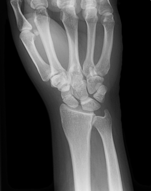
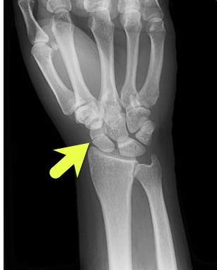
25 year-old female presents following a fall from her horse 5 days ago. She complains of severe pain in the radial aspect of her right wrist and has no other injuries.  Plain films at an outside ED immediately following the injury were negative, and repeat plain films are shown here. What’s the diagnosis?

Answer: Non-displaced Scaphoid Waist Fracture

  • The most common carpal bone fracture (60-70% of all carpal fractures). 10-30% of scaphoid fractures are not detected on the first set of plain films, but “scaphoid view” plain films (i.e., AP wrist with ulnar deviation) can improve the view of the scaphoid.
  • If a scaphoid fracture is clinically suspected, the patient should be placed in a thumb spica splint and follow up in 7-10 days for repeat plain films and reexamination
  • The most feared complication of a scaphoid fracture is avascular necrosis (AVN) of the proximal fracture segment. AVN is more likely in unstable scaphoid fractures, for example those that are proximal, oblique, displaced >1 mm, rotated, or comminuted. These require surgical consult and long arm thumb spica splint.
  • Stable fractures can be splinted with a short arm thumb spica splint and patients should be instructed to follow up with orthopedics in 7-10 days

References:

Escarza, Robert et al. “Chapter 266. Wrist Injuries.” Tintinalli’s Emergency Medicine a Comprehensive Study Guide, 7e.  Eds, Judith E. Tintinalli, et al.  New York, NY: McGraw-Hill, 2011.

DeAngelis, Michael A and David A Wald. “Wrist.” Simon’s Emergency Orthopedics, 7e.  Ed. Scott C Sherman.  New York, NY: McGraw-Hill, 2014

Jordanov, Martin I and Robert Warne Fitch. “Chapter 9 Upper Extremity.” The Atlas of Emergency Radiology. Eds. Jake Block et al.  New York, NY: McGraw-Hill, 2013

Wednesday Image Review

What’s the Diagnosis? By Dr. Edward Guo

A 20 year old male presents to the emergency department via EMS for left knee pain. He was playing basketball when he jumped and felt a “pop” in his left knee and has been unable to walk on his left leg since. He denies falling. On exam, the left lower extremity is distally neurovascularly intact with normal strength, sensation, and a palpable pulse. There is slight bogginess and swelling with tenderness to palpation to the inferior knee. He is unable to extend at the knee. A point of care ultrasound of the bilateral knees is performed and shown below. What’s the diagnosis?

Answer: Left patellar tendon rupture

  • Commonly occurs from forced quadriceps contraction or falling on a flexed knee.
  • Associated with a high-riding patella also known as patella alta which can be appreciated on physical exam and lateral radiographs of the knee.
  • There is emerging data demonstrating point of care ultrasound as a quick and effective method to diagnose tendon injuries in the emergency department compared to physical exam, x-ray imaging, and MRI.
  • Treatment:
    • Incomplete tears with intact extensor mechanism can be immobilized and followed up outpatient with orthopedics.
    • Complete tears or loss of extensor mechanism should prompt orthopedic consultation in the ED as expedited surgical repair is often indicated.

References:

Bengtzen R. Knee Injuries. In: Tintinalli JE, Ma O, Yealy DM, Meckler GD, Stapczynski J, Cline DM, Thomas SH. eds. Tintinalli’s Emergency Medicine: A Comprehensive Study Guide, 9e. McGraw-Hill Education; 2020.

Berg, K., Peck, J., Boulger, C., & Bahner, D. P. (2013). Patellar tendon rupture: an ultrasound case report. BMJ case reports2013, bcr2012008189. https://doi.org/10.1136/bcr-2012-008189

Wu TS, Roque PJ, Green J, et al. Bedside ultrasound evaluation of tendon injuries. Am J Emerg Med. 2012;30(8):1617-1621. doi:10.1016/j.ajem.2011.11.004

Tuesday Advanced Cases

Alcohol Withdrawal By Dr. Sandhya Ashokkumar

HPI

  • A 53-year-old male with a history of chronic daily alcohol use presents to the ED via EMS after a witnessed seizure at home
  • The medics say he drinks every day, but his last drink was 3 days ago because he was not able to go to the store (history obtained from the sister who witnessed the seizure)

Physical Examination

T 98.3F, BP 177/106, HR 191, RR 22, Sat 93% 

  • Patient appears anxious, uncomfortable, and is actively vomiting blood tinged sputum
  • He appears confused and is not answering questions appropriately
  • He is tachycardic and his lungs are clear to auscultation bilaterally
  • Abd: soft NTND

Differential 

  • Alcohol withdrawal/ Delirium Tremens
  • Thyrotoxicosis
  • Sepsis
  • Pulmonary embolism
  • Heart failure

Workup and Management

  • The nurse informs you that the patient is seizing
  • This patient is exhibiting evidence of delirium tremens (psychomotor agitation and autonomic instability) and alcohol withdrawal seizure
  • The patient is no longer tolerating his secretions, he is confused, gurgling, and requires a definitive airway
  • After intubation, you bolus the patient with propofol and start him a propofol infusion
  • The patient is admitted to the ICU for further management

Teaching Points

  • ETOH withdrawal begins 6-8 hours after last intake and peaks in 72 hours 
  • Symptom based treatment via the CIWA score can help stratify patients, scores >15 indicate severe withdrawal
  • Start with IV diazepam at 10mg or lorazepam at 4mg and repeat them in doubling doses
  • Consider adding phenobarbital for refractory cases (i.e. after 200 mg of diazepam)
  • Consider propofol as the induction agent and sedative post-intubation as propofol potentiates GABA receptor activity and inhibits NMDA receptors Summary 
  • Delerium tremens can result in death from hyperthermia, arrhythmia and seizures

Sources:

Long D, Long B, Koyfman A. The emergency medicine management of severe alcohol withdrawal. The American Journal of Emergency Medicine. 2017;35(7):1005-1011. doi:10.1016/j.ajem.2017.02.002

Schuckit MA, Author Affiliations From the Department of Psychiatry. Recognition and Management of Withdrawal Delirium (Delirium Tremens): NEJM. New England Journal of Medicine. https://www.nejm.org/doi/10.1056/NEJMra1407298. Published February 5, 2015. Accessed December 9, 2020.

Wednesday Image Review

What’s the Diagnosis? By Dr. Edward Guo

A 40 year old female presents to the emergency department via EMS for shortness of breath. Prior to arrival to the ED, the patient was hypoxic and in severe respiratory distress with absent left lung sounds prompting needle thoracostomy and rapid sequence intubation by EMS. Vital signs are BP 108/70, HR 102, Temp 98F, RR 16, SpO2 99% on 50% FiO2. A left sided chest tube is placed without complication. Chest x-ray confirms appropriate positioning of the endotracheal tube and chest tube with expansion of the left lung. Four hours later, the ventilator is alarming due to elevated peak and plateau pressures. SpO2 is 90%. There is no change with suctioning. A new chest x-ray is obtained and is shown below. What’s the diagnosis?

Answer: Reexpansion pulmonary edema

  • Reexpansion pulmonary edema is a rare but potentially fatal complication following drainage of a pneumothorax or pleural effusion. The pathophysiology is poorly understood but is thought to involve an inflammatory response leading to increased pulmonary capillary permeability.
  • Risk factors include large size pneumothorax, large volume pleural effusion, rapid reexpansion, and prolonged duration of symptoms (> 72 hours).
    • Prevention includes limiting drainage of pleural effusions to a maximum volume of 1.5 liters in one attempt.
  • Imaging will demonstrate unilateral airspace opacities in portions of the lung that were previously collapsed.
  • Treatment is supportive with supplemental oxygen and observation. Most patients recover without adverse outcomes.

References:

Nicks BA, Manthey DE. Pneumothorax. In: Tintinalli JE, Ma O, Yealy DM, Meckler GD, Stapczynski J, Cline DM, Thomas SH. eds. Tintinalli’s Emergency Medicine: A Comprehensive Study Guide, 9e. McGraw-Hill Education; 2020.

Asciak R, Bedawi EO, Bhatnagar R, et al British Thoracic Society Clinical Statement on pleural procedures Thorax 2023;78:s43-s68.

Morioka H, Takada K, Matsumoto S, Kojima E, Iwata S, Okachi S. Re-expansion pulmonary edema: evaluation of risk factors in 173 episodes of spontaneous pneumothorax. Respir Investig. 2013;51(1):35-39. doi:10.1016/j.resinv.2012.09.003

https://radiopaedia.org/articles/re-expansion-pulmonary-oedema

Tuesday Advanced Cases

Hypothermia Arrhythmia by Dr. Edward Guo

Case: A 29 year old male with a past medical history of polysubstance use presents to the ED in December via EMS for a suspected overdose. History is limited due to patient cooperation. EMS states that he was found outside in a puddle, minimally responsive. He was given 2mg IM naloxone by EMS and became acutely agitated and combative afterward, requiring 5mg IM midazolam and 5mg IM haloperidol upon arrival. Fingerstick glucose 226. EKG is obtained and shown below.

Exam: BP 182/84, HR 111, T 86.1F, RR 18, SpO2 100%
Disheveled appearing male in wet clothes, intermittently thrashing. Cold to touch. Pupils 5mm bilaterally. No signs of trauma. GCS E3 V2 M5. Moves all extremities equally. Heart rate is tachycardic and irregular.

EKG interpretation: atrial fibrillation with rapid ventricular response with Osborn waves

Differential diagnosis: polysubstance use, environmental cold exposure, severe sepsis, hypothyroidism

Case continued: Active rewarming is initiated by removing wet clothes, administering warmed IV fluids, and placing a bair hugger. Labs are notable for a creatinine kinase of 3966. The patient’s temperature, heart rate, and mental status significantly improve within 5 hours, and his repeat EKG shows normal sinus rhythm without Osborn waves. He is ultimately admitted to medicine.

Pearls:

  • The cardiovascular response to cold is peripheral vasoconstriction and initial increase in heart rate and blood pressure. As core temperature drops below 32C, there is myocardial irritability and risk of cardiovascular collapse.
    • Atrial fibrillation and flutterare common arrhythmias associated with hypothermia.
    • Rescue collapse is a term to describe cardiac arrest that occurs during extrication or transport of a profoundly hypothermic patient due to profound myocardial irritability.
  • Osborn waves are positive deflections at the end of the QRS complex that are non-specific but may occur in temperatures below 32C.
    • Size of the wave correlates with the degree of hypothermia but has no prognostic value.
  • As temperature continues to drop, EKG changes are variable but classically include bradycardia with prolonged PR, QRS, and QTc. Heart block or ventricular dysrhythmias may be encountered as well. Asystole is the common final dysrhythmia.
  • Rewarming is the treatment of choice.
    • Atrial dysrhythmias such as atrial fibrillation will often resolve with warming.
    • Cardioversion for unstable arrhythmias should be attempted but may be refractory in severe hypothermia.

References:

Brown DA. Hypothermia. In: Tintinalli JE, Ma O, Yealy DM, Meckler GD, Stapczynski J, Cline DM, Thomas SH. eds. Tintinalli’s Emergency Medicine: A Comprehensive Study Guide, 9e. McGraw-Hill Education; 2020.

Hoek T. 2010 American Heart Association Guidelines for Cardiopulmonary Resuscitation and Emergency Cardiovascular Care. Circulation. 2010. 122:5829-5861

Wednesday Image Review

What’s the Diagnosis? By Dr. Edward Guo

A 50 year old male with a past medical history of Crohn disease with ileocolectomy presents via EMS for shortness of breath. Prior to arrival to ED, patient was found to be hypoxic and in acute respiratory distress prompting rapid sequence intubation by EMS. Vital signs are notable for hypotension and tachycardia. On exam, there are equal breath sounds bilaterally. His abdomen is distended with bruising on the left flank. GCS is 3T. A portable chest x-ray is obtained to confirm endotracheal tube placement and is shown below. What’s the diagnosis?

Answer: Pneumoperitoneum

  • Most commonly caused by gastrointestinal perforation from etiologies such as peptic ulcer disease, traumatic injury, bowel obstruction, or infection.
  • While CT is the gold standard for diagnosis, a chest x-ray may be utilized to quickly assess for presence of subdiaphragmatic air.
    • Sensitivity of upright chest x-ray to detect pneumoperitoenum varies across studies but is up to 80%.
    • Upright positioning for 10 minutes prior to radiograph or lateral upright positioning may increase sensitivity to over 90%.
    • Specificity is approximately 90%.
  • Management includes emergent surgical consultation, broad spectrum antibiotics with anaerobic coverage, and gastric decompression.

References:

Bogle AM, Gratton MC. Peptic Ulcer Disease and Gastritis. In: Tintinalli JE, Ma O, Yealy DM, Meckler GD, Stapczynski J, Cline DM, Thomas SH. eds. Tintinalli’s Emergency Medicine: A Comprehensive Study Guide, 9e. McGraw-Hill Education; 2020.

Stapakis JC, Thickman D. Diagnosis of pneumoperitoneum: abdominal CT vs. upright chest film. J Comput Assist Tomogr 1992;16:713–16.

Woodring JH, Heiser MJ. Detection of pneumoperitoneum on chest radiographs: comparison of upright lateral and posteroanterior projections. Am J Roentgenol 1995;165:45–7.

Wednesday Image Review

What’s the Diagnosis? By Dr. Katie Selman

A 63 year old female is brought in by EMS after being found down. She has multiple ecchymoses on her chest and bilateral flanks. GCS is 6. After intubation, she is taken for CT head/cervical spine and a CT chest/abdomen/pelvis with contrast. Upon return from CT, x-rays are done (shown below) to further evaluate bruising and a laceration to her L elbow. What’s the diagnosis?

Answer: Contrast extravasation

  • Predisposing factors for contrast extravasation
    • Small IV gauge (22G or less)
    • More distal access (hand)
    • Rapid injection of contrast
  • Incidence: up to 1% of patient receiving IV contrast through peripheral IV
  • Most common symptoms: local pain, swelling
  • Complications occur in < 1 %  (more common with large volume and in patients with atherosclerosis, venous insufficiency, or impaired lymphatic drainage)
    • Compartment syndrome
    • Tissue necrosis
  • Close monitoring required following extravasation
    • Compartment checks, vascular checks, and monitoring of overlying skin
    • Surgery consult for any signs of compartment syndrome or tissue injury
    • Elevate limb, warm compresses may be used
    • Patients rarely require more than conservative supportive treatment

References:

Sbitany, H., Koltz, P. F., Mays, C., Girotto, J. A., & Langstein, H. N. (2010). CT contrast extravasation in the upper extremity: Strategies for management. International Journal of Surgery, 8(5), 384-386. doi:10.1016/j.ijsu.2010.06.002

Sonis, J. D., et al (2018). Implications of iodinated contrast media extravasation in the emergency department. The American Journal of Emergency Medicine, 36(2), 294-296. doi:http://dx.doi.org/10.1016/j.ajem.2017.11.012