HPI: Pt is a 41 y/o male who presents to the ED w/ significant wrist pain s/p MVC. He was the unrestrained driver of a car, T boned at a moderate speed, + broken windshield, + airbag deployment, – entrapment. R wrist is swollen tender over the mid dorsal aspect w/o any open lacerations.
Vitals: HR 101, BP 121/91, Temp 98F, RR 15
Physical Exam: GCS 15 Cardiac, Pulm & Abd unremarkable.
Extremities: LUE, LLE, RLE – unremarkable. RUE: Strength 5/5 throughout, sensation intact at C5-T1 and distally, able to make an “okay”, “thumbs up” sign. Point tenderness over R dorsal mid wrist and snuff box w/o deformity or skin changes, + swelling, compartments soft. Limitation w/ flexion/extension of the R wrist, normal finger cascade when closing hand,
Differential: Scaphoid fracture, scapholunate dissociation, proximal MCP fracture, distal radius or ulnar fracture, other carpal injury.
Xray
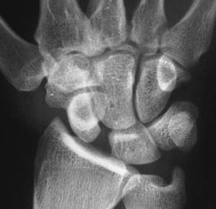
Scapholunate Dissociation is a common result of hand trauma and it may be acute (traumatic) or chronic (in the setting of degenerative injuries). Often associated w/ intraarticular distal radius and other carpal fractures. Mechanism typically involves forced wrist extension and ulnar deviation. Think FOOSH injury.
Associated injury often is a “DISI” – Dorsal Intercalated Segmental Instability. A scapholunate dissociation causes the scaphoid bone to rotate anterior while lunate rotates posteriorly – if untreated can progress to chronic degenerative arthritis.
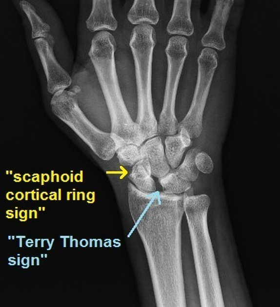
Diagnosis: XR AP radiograph: > 3mm of widening between the scaphoid and lunate on a “clenched fist view”.
Signet Ring Sign: indicative of a scapholunate dissociation. As the scaphoid rotates the scaphoid tubercle appears more rounded. Can also be seen in perilunate and lunate dislocations.
Treatment: Pain control, splinting (sugar tong or thumb spica) and repeat imaging in 1-2 weeks w/ ortho followup. Often requires operative fixation in outpt setting. If associated w/ perilunate, lunate, scaphoid dislocation – requires urgent reduction prior discharge.
Complications: Chronic pain and degenerative disease of associated structures, neurovascular compromise, joint instability
References:
1. Stevenson M, Levis JT. Image diagnosis: Scapholunate dissociation. The Permanente journal. 2019. Accessed January 14, 2025. https://pmc.ncbi.nlm.nih.gov/articles/PMC6443367/#f1-18-237.
2. Scapholunate ligament tear – cortical ring sign | radiology case | radiopaedia.org. Accessed January 15, 2025. https://radiopaedia.org/cases/scapholunate-ligament-tear-cortical-ring-sign.
3. Scapholunate Ligament Injury & Disi. Orthobullets. Accessed January 14, 2025. https://www.orthobullets.com/hand/6041/scapholunate-ligament-injury-and-disi.
4. Rap corependium. EM. Accessed January 14, 2025. https://www.emrap.org/corependium/chapter/recrf1uDQslSAzIEL/Wrist-Dislocations#h.t4rehgq0mdhi.