Friday Board Review

Board Review with Dr. Edward Guo

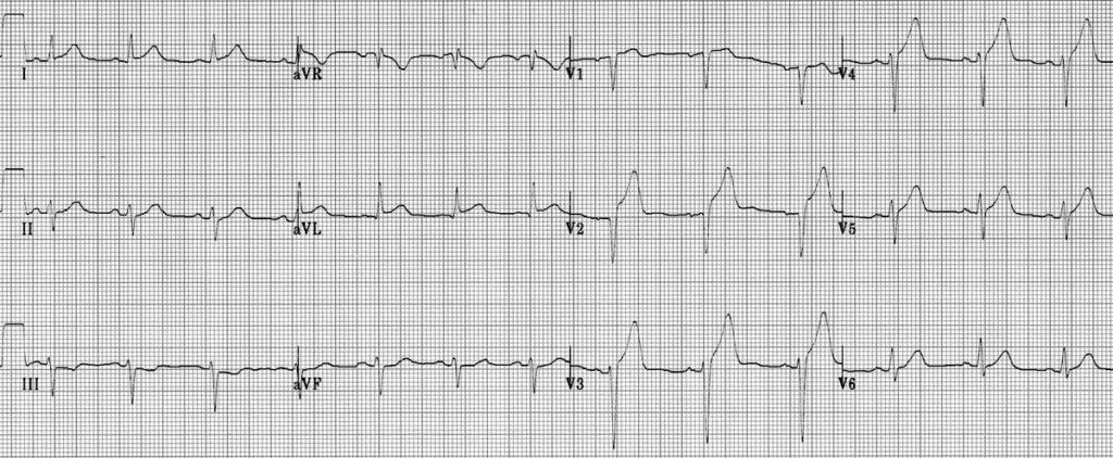
A 68 year old male with a history of hypertension and diabetes presents with chest pain. He was working in his yard when he suddenly felt severe, pressure across his chest. Vital signs are: Temp 98.8, HR 80, BP 162/100, RR 20, SpO2 95% RA. He appears in moderate distress and is diaphoretic. His EKG is shown below. Which of the following is NOT recommended for management of this patient?

ECG Anterolateral AMI STEMI 2

A: activation of the cath lab

B: aspirin

C: clopidogrel

D: nasal cannula oxygen

E: nitroglycerin

Answer: nasal cannula oxygen

This patient is experiencing a STEMI. His infarct is likely anteroseptal as evidenced by ST segment elevations and hyperacute T waves in V2-V4 as well as ST segment elevation in I and aVL with reciprocal ST segment depressions in III and aVF. The Air Versus Oxygen In ST-Segment-Elevation MyocarDial Infarction (AVOID) trial conducted in 2015 demonstrated that supplemental oxygen in STEMI patients without hypoxia may increase early myocardial injury and was associated with larger myocardial infarct size at 6 months. Activation of the cath lab and loading with aspirin and clopidogrel are recommended. Nitroglycerin may provide symptomatic relief for this patient but should be used cautiously in patients with evidence of right ventricular infarction. 

References:

Stub, D., Smith, K., Bernard, S., Nehme, Z., Stephenson, M., Bray, J. E., Cameron, P., Barger, B., Ellims, A. H., Taylor, A. J., Meredith, I. T., Kaye, D. M., & AVOID Investigators (2015). Air Versus Oxygen in ST-Segment-Elevation Myocardial Infarction. Circulation131(24), 2143–2150. https://doi.org/10.1161/CIRCULATIONAHA.114.014494

Wednesday Image Review

From the EMDaily Archives: What’s the Diagnosis? By Dr. Abby Renko

A 45 year old male with a history of ESRD on hemodialysis and insulin dependent diabetes presents with left knee pain after slipping at work. Vitals are within normal limits. On exam, the left lower extremity is distally neurovascularly intact with palpable distal pulses. There is obvious swelling over the knee with the worst pain superior to the patella. He is unable to extend the lower leg. An x-ray is performed and shown below. What’s the diagnosis?

Answer: quadriceps tendon rupture

  • At first glance, lateral XR does not show obvious deformity… however, you may notice calcifications just superior to the patella representing retracted tendon.
  • Quadriceps tendon ruptures tend to occur in individuals > 40, while patellar tendon ruptures occur more frequently in individuals < 40.
    • Overall, quadricep tendon ruptures are more common (risk factors include rheumatologic disease, renal failure, DM, chronic steroid use).
  • In both injuries, classic exam finding is inability to extend the knee. You can often palpate a defect just above the patella in quadriceps tendon ruptures.
  • High riding patella (“patella alta”) on lateral films is more frequently seen in patellar tendon ruptures. Low riding patella (“patella baja”) may be seen on lateral film with complete quadricep tendon tear (intact patellar tendon displaces patella inferiorly).
  • X-rays may be normal! Ultrasound is the diagnostic modality of choice in the ED as it has both high sensitivity and specificity.
  • Orthopedic consultation is always warranted for these cases. Management involves a knee immobilizer and prompt follow up. Early surgical repair is associated with better outcomes in severe cases.

References:

Bengtzen RR, Glaspy JN, Steele MT. Knee Injuries. In: Tintinalli JE, Stapczynski JS, et al., eds. Tintinalli’s Emergency Medicine: A Comprehensive Study Guide. 8th ed. New York, NY: McGraw-Hill; 2016: (Ch) 274. 

Pope JD, El Bitar Y, Mabrouk A, et al. Quadriceps Tendon Rupture. [Updated 2023 Apr 22]. In: StatPearls [Internet]. Treasure Island (FL): StatPearls Publishing; 2023 Jan-. Available from: https://www.ncbi.nlm.nih.gov/books/NBK482389/

Tuesday Advanced Cases & Procedure Pearls

Advanced Cases: Chest Pain with Dr. Erica Westlake

A 25 year old female with a history of lupus presents with chest pain and shortness of breath. Her vitals are significant for tachycardia and are otherwise within normal limits. Her initial EKG is shown below:

The STEMI phone is contacted. Prior to cath lab activation, cardiology recommends a bedside echocardiogram which demonstrates the following:

Cardiology performed echocardiogram re-demonstrates a circumferential pericardial effusion without regional wall motion abnormalities or tamponade physiology. The cath lab was not activated, there was no elevation in troponins, and the patient was ultimately diagnosed with acute pericarditis.

Diagnostic Criteria for Acute Pericarditis (requires 2 of the following)
Characteristic chest pain (sharp or pleuritic, improved sitting up and leaning forward, worse lying down)
Pericardial friction rub
New or worsening pericardial effusion
Suggestive EKG changes (diffuse ST segment elevations)
  • Spontaneous Coronary Artery Dissection (SCAD) is a life threatening process involving a tear within the coronary arterial wall.
  • SCAD predominantly affects young (< 50) women especially during pregnancy and post-partum periods. It can appear identical to a STEMI on EKG despite “non-occluded” coronary vessels. It is commonly associated with a rise in cardiac enzymes and regional wall motion abnormalities on echocardiogram. It is definitively diagnosed via coronary angiography. Treatment will vary based on high vs low risk features.

References:

Diercks DB, Hollander JE, Chang A. Acute Coronary Syndromes. In: Tintinalli JE, Ma O, Yealy DM, Meckler GD, Stapczynski J, Cline DM, Thomas SH. eds. Tintinalli’s Emergency Medicine: A Comprehensive Study Guide, 9e. McGraw Hill; 2020.

Kim ESH. Spontaneous Coronary-Artery Dissection. N Engl J Med. 2020 Dec 10;383(24):2358-2370. 

Ornato, J. P., Ou, N., Radford, M. J., Tamis-Holland, J. E., Tommaso, C. L., … Zhao, D. X. (2013). 2013 ACCF/AHA guideline for the management of ST-elevation myocardial infarction: a report of the American College of Cardiology Foundation/American Heart Association Task Force on Practice Guidelines. Journal of the American College of Cardiology61(4), e78–e140. https://doi.org/10.1016/j.jacc.2012.11.019

Whelan LJ. Comorbid Disorders in Pregnancy. In: Tintinalli JE, Ma O, Yealy DM, Meckler GD, Stapczynski J, Cline DM, Thomas SH. eds. Tintinalli’s Emergency Medicine: A Comprehensive Study Guide, 9e. McGraw Hill; 2020.

Friday Board Review

Board Review by Dr. Edward Guo

A 3 month old male born full term with no complications presents for a right groin mass. His mother first noticed it two days ago when he cried. Otherwise, he has been acting normally, drinking formula regularly, and having several bowel movements daily. Vital signs and weight are within normal limits for age. Exam shows a happy appearing infant that cries when taken away from his mother. When he cries, a 3 cm soft, non-tender mass is appreciated in the right inguinal region that is easily reducible. There is no overlying erythema or skin changes. Genital exam reveals palpable bilateral testes with no additional masses in the scrotum. Which of the following is the most appropriate management of this condition?

A: admit for observation

B: discharge with expedited surgical follow up

C: discharge with reassurance that it will likely spontaneously resolve

D: emergent surgical consultation

Answer: discharge with expedited surgical follow up

This infant is presenting with a reducible inguinal hernia that increases in size with increased intra-abdominal pressure such as with crying or straining. It is most common in premature males in the first year of life. Unlike in adults, children require surgical referral for interval repair of simple reducible inguinal hernias due to the higher risk of incarceration. Thus, reassurance that it will likely spontaneously resolve without surgical follow up is incorrect. If the hernia was incarcerated, emergent surgical consultation with admission for prompt surgical repair would be appropriate management. 

References:

Fleischman R.J., & Meckler G (2020). Acute abdominal pain in infants and children. Tintinalli J.E., & Ma O, & Yealy D.M., & Meckler G.D., & Stapczynski J, & Cline D.M., & Thomas S.H.(Eds.), Tintinalli’s Emergency Medicine: A Comprehensive Study Guide, 9e. McGraw Hill.

Wednesday Image Review

What’s the Diagnosis? By Dr. Edward Guo

A 63 year old male presents for “floaters” in his right eye for two weeks. He wears reading glasses at baseline. He denies pain or known injury to the eye. On exam, his visual acuity is 20/30 OD, 20/20 OS corrected with reading glasses. Pupils are equal, round, and reactive to light. IOP is 8 OD, 9 OS. There are no areas of focal uptake with fluorescein stain. POCUS of the right orbit is shown below. What’s the diagnosis?

Answer: Posterior Vitreous Detachment and Vitreous Hemorrhage (bright echogenic membrane horizontally across the posterior chamber not attached at the optic nerve and multiple free-flowing areas of varying hyperechogenicity that are mobile with eye movement)

  • Presentation may vary from sudden onset floaters and generalized hazy vision to complete vision loss depending on severity
  • May sometimes be a precursor to a retinal detachment
  • Important to distinguish from a retinal detachment which will demonstrate a “V” shaped echogenic membrane attached to the optic nerve on POCUS
  • Management is ophthalmology consultation especially if retinal detachment is suspected as it is a true ophthalmologic emergency

References:

Walker R.A., & Adhikari S (2020). Eye emergencies. Tintinalli J.E., & Ma O, & Yealy D.M., & Meckler G.D., & Stapczynski J, & Cline D.M., & Thomas S.H.(Eds.), Tintinalli’s Emergency Medicine: A Comprehensive Study Guide, 9e. McGraw Hill. 

Lahham, S., Ali, Q., Palileo, B. M., Lee, C., & Fox, J. C. (2019). Role Of Point Of Care Ultrasound In The Diagnosis Of Retinal Detachment In The Emergency Department. Open access emergency medicine : OAEM11, 265–270. https://doi.org/10.2147/OAEM.S219333

www.emra.org/emresident/article/floaters-retinal-detachment-posterior-vitreous-detachment-or-vitreous-hemorrhage

Friday Board Review

Board Review by Dr. Edward Guo

A 62-year old male with a past medical history of hypertension, hyperlipidemia, and coronary artery disease presents with chest pain. He was discharged from the hospital 2 days ago after being treated for a STEMI which occurred 5 days ago. The pain is mid-sternal, worse with deep inspiration and lying flat. He has been taking all of his medications including aspirin and clopidogrel as instructed. Vital signs are: BP 142/76, HR 102, T 37.2 C, RR 18, SpO2 96%. On exam, he is sitting upright and appears in mild discomfort but otherwise has no focal findings. EKG shows Q waves in II, III, and aVF without significant other changes. POCUS reveals a small circumferential pericardial effusion with a normal ejection fraction. Which of the following is the first line treatment of this patient’s suspected condition?

A: colchicine

B: heparin

C: high dose aspirin

D: naproxen

E: prednisone

Answer: high dose aspirin

This patient is presenting with peri-infarction pericarditis which is an uncommon complication of STEMIs due to the increasing use of reperfusion therapy. Despite not having classic EKG changes suggestive of pericarditis, he meets diagnostic criteria for acute pericarditis. According to the American College of Cardiology and American Heart Association, high dose aspirin (650 mg every 6 hours) is the first line treatment of acute pericarditis after STEMI (Class I recommendation). In contrast to viral, idiopathic, or autoimmune causes of pericarditis, NSAIDs such as naproxen or glucocorticoids are not recommended due to potential for myocardial scar thinning and infarct expansion. Colchicine may be used as an adjunctive medication if high dose aspirin is not effective. Heparin is not recommended for the treatment of acute pericarditis. 

Diagnostic Criteria for Acute Pericarditis (requires 2 of the following)
Characteristic chest pain (sharp or pleuritic, improved sitting up and leaning forward, worse lying down)
Pericardial friction rub
New or worsening pericardial effusion
Suggestive EKG changes (diffuse ST segment elevations)

References:

O’Gara, P. T., Kushner, F. G., Ascheim, D. D., Casey, D. E., Jr, Chung, M. K., de Lemos, J. A., Ettinger, S. M., Fang, J. C., Fesmire, F. M., Franklin, B. A., Granger, C. B., Krumholz, H. M., Linderbaum, J. A., Morrow, D. A., Newby, L. K.,

Ornato, J. P., Ou, N., Radford, M. J., Tamis-Holland, J. E., Tommaso, C. L., … Zhao, D. X. (2013). 2013 ACCF/AHA guideline for the management of ST-elevation myocardial infarction: a report of the American College of Cardiology Foundation/American Heart Association Task Force on Practice Guidelines. Journal of the American College of Cardiology61(4), e78–e140. https://doi.org/10.1016/j.jacc.2012.11.019

Friday Board Review

Board Review with Dr. Edward Guo

An 18 year old male with a history of type 1 diabetes mellitus presents for a foot wound. He was barefoot playing soccer at a park when he suddenly felt sharp pain on the sole of his right foot and saw a metal nail in the grass. Vital signs are within normal limits. POC glucose is 140. The extremity is neurovascularly intact and shows a subcentimeter puncture wound on the plantar surface with no surrounding erythema or active bleeding. The area is tender to palpation but he is able to ambulate with minimal pain. His tetanus is up to date. In addition to irrigation of the wound, which of the following is most appropriate management for this patient?

A: administer a five-day course of cephalexin

B: administer a five-day course of ciprofloxacin

C: close the wound and discharge the patient

D: discharge with primary care follow up

Answer: administer a five-day course of cephalexin

Antibiotic prophylaxis is recommended for puncture wounds with high-risk features including plantar punctures, bite wounds, heavy contamination, or patients with diabetes or immunosuppression. Most soft tissue infections from puncture wounds are caused by gram-positive organisms. Thus, cephalexin is the most appropriate option listed. Ciprofloxacin would be appropriate if the patient suffered a puncture wound through a shoe as it is thought that pseudomonas colonizes the foam soles. It is generally not recommended to close high risk wounds due to increased risk of infection.

Osteomyelitis
OrganismAssociation
Staphylococcus aureusMost common overall
Salmonella sp.Sickle cell disease
Pseudomonas sp.Puncture through shoe sole
Pasteurella multocidaDog and cat bites

References:

Quinn J (2020). Puncture wounds and bites. Tintinalli J.E., & Ma O, & Yealy D.M., & Meckler G.D., & Stapczynski J, & Cline D.M., & Thomas S.H.(Eds.), Tintinalli’s Emergency Medicine: A Comprehensive Study Guide, 9e. McGraw Hill.