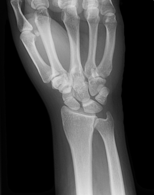
A 38 year old male presents to the ED with a chief complaint of right wrist pain that began after a fall off a motorcycle the day prior. On exam, the patient is noted to have tenderness along his right distal radius, snuffbox tenderness, wrist swelling, and is unable to flex/extend the wrist. Normal pulses and sensation is present.
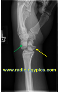
Wrist X-rays demonstrate the following:
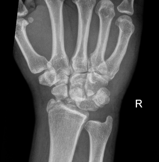
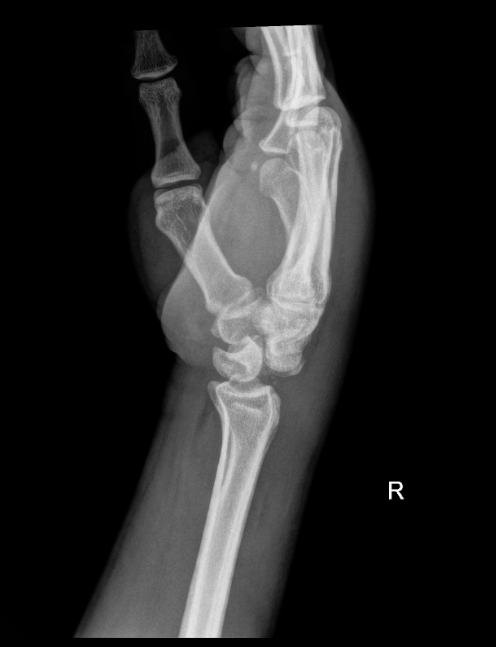
What is the diagnosis?
Perilunate dislocation, scaphoid fracture, radial styloid fracture
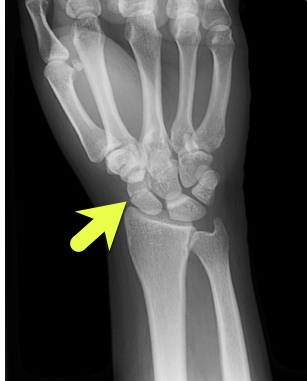
Perilunate Dislocation:
- Occur due to a high energy traumatic injury
- Multiple wrist ligaments are injured with resultant dislocation of the capitate dorsally
- Often associated with fractures of the radius, ulnar, or carpal bones
- Imaging:
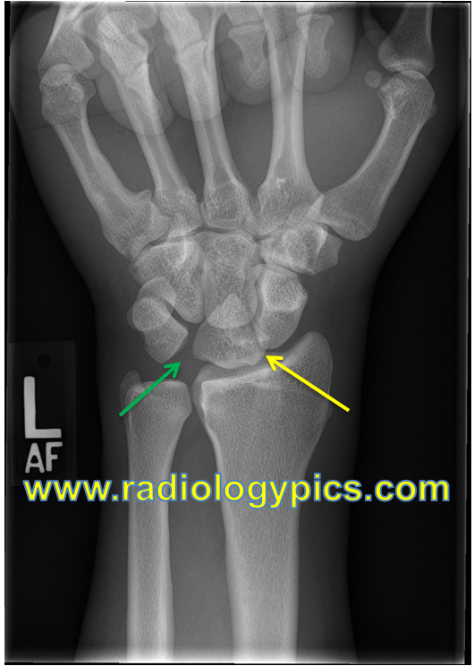
- Lunate stays in place, dislocated bone is actually the capitate!
- AP/PA XR may demonstrate “piece-of-pie” sign: triangular appearance of the lunate (yellow arrow)
- Lateral XR: Proximal and dorsal displacement of the capitate (yellow arrow) with volar displacement of the lunate (green arrow).
- The lunate remains articulated with the radius differentiating it from a lunate dislocation (lunate dislocation would have a “spilled-teacup” sign)
- Management:
- Emergent closed reduction is indicated to minimize complications such as: median nerve injury, cartilage damage, wrist function issues
- Sugar-tong splint
- Urgent orthopedic follow-up as most will require surgical fixation
References:
Cheffers M. Wrist Reduction Techniques. In: Johnson W, Nordt S, Mattu A and Swadron S, eds. CorePendium. Burbank, CA: CorePendium, LLC. https://www.emrap.org/corependium/chapter/recoPqKOBgkCSHesR/Wrist-Reduction-Techniques#h.493xci6kkby6. Updated December 21, 2022. Accessed August 15, 2024.
Mark Karadsheh. “Lunate Dislocation (Perilunate Dissociation).” Orthobullets, 5 Nov. 2022, www.orthobullets.com/hand/6045/lunate-dislocation-perilunate-dissociation. “Solution to Unknown Case #30 – Perilunate Dislocation.” RADIOLOGYPICS.COM, 6 Jan. 2014, radiologypics.com/2013/03/28/perilunate-dislocation/.