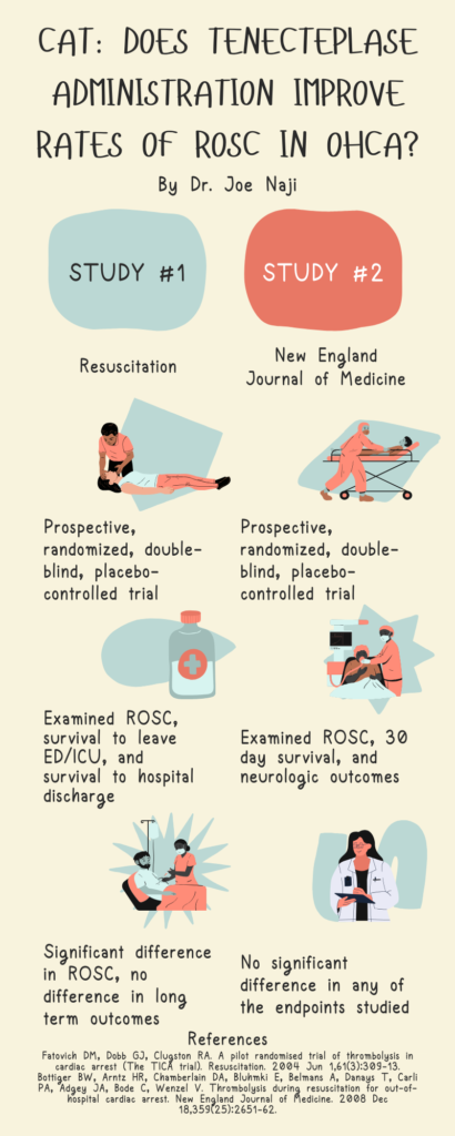
CAT: Does Tenecteplase administration improve rates of ROSC in OHCA? By Dr. Joe Naji


A 33 year old male with no past medical history presents for right hand pain. He works in construction. A few weeks ago, there was a wooden splinter in his palm that has grown into a nodule. He denies any drug use. Vital signs are within normal limits. The right upper extremity is neurovascularly intact with full range of motion. There is a 1.5 cm pedunculate lesion shown below. It is firm and minimally tender to palpation with no bleeding or drainage. Which of the following is the appropriate treatment?

A: discharge with dermatology follow up
D: incision and drainage
C: oral cephalexin
D: topical mupirocin ointment
Answer: A. discharge with dermatology follow up
This patient is presenting with a pyogenic granuloma, a benign vascular tumor that classically occurs after minor trauma in young adults and pregnant women. It most commonly occurs in the hands or oral cavity and will recur without proper treatment. A dermatologist can confirm the diagnosis with a biopsy. Incision and drainage, oral cephalexin, or topical mupirocin ointment are useful in the management of abscesses and infected wounds but are not appropriate for a pyogenic granuloma. Definitive treatment includes surgical excision, laser therapy, or electrocautery.
References:
Holahan H, & Morrell D.S., & McShane D.B. (2020). Skin disorders: extremities. Tintinalli J.E., & Ma O, & Yealy D.M., & Meckler G.D., & Stapczynski J, & Cline D.M., & Thomas S.H.(Eds.), Tintinalli’s Emergency Medicine: A Comprehensive Study Guide, 9e. McGraw Hill.
A 17 year old male presents 20 minutes after having his tooth knocked out during a hockey game brawl. His parents preserved the tooth wrapped in a dry paper towel. He denies loss of consciousness or vomiting. Vitals are within normal limits. Exam shows a well-appearing male in no apparent distress with loss of tooth #8. The tooth socket is hemostatic, and there is no deformity or tenderness to palpation. The tooth is irrigated, replanted, and splinted. Which of the following is indicated at this time?
| A | calcium hydroxide paste |
| B | consult oral maxillofacial surgery |
| C | CT facial bones |
| D | doxycycline |
D. Doxycycline
This patient experienced a tooth avulsion with subsequent ED replantation. Notably, time to replantation is the most important prognostic factor. Doxycycline has also demonstrated some benefit in successful replantation of the tooth. Calcium hydroxide paste is used in dental fractures, not avulsions. Consulting oral maxillofacial surgery is not necessary after ED replantation, but the patient should have expedited dental follow up. CT facial bones will unlikely show an acute fracture given the patient has no clinical findings to suggest injury. A panoramic x-ray may be beneficial in confirming tooth position after replantation.
| Tooth Avulsion |
| Time is tooth! Rule of thumb: each minute = 1% lower chance of successful replantation Transport solutions: Hank solution > milk > saliva > saline Handle by crown, irrigate gently with saline (do not disrupt periodontal ligament fibers) Pain control, manual pressure, splint Doxycycline Soft diet Urgent dental follow up |
References:
Beaudreau R.W. (2020). Oral and dental emergencies. Tintinalli J.E., & Ma O, & Yealy D.M., & Meckler G.D., & Stapczynski J, & Cline D.M., & Thomas S.H.(Eds.), Tintinalli’s Emergency Medicine: A Comprehensive Study Guide, 9e. McGraw Hill.
Amsterdam JT. Oral medicine. In: Marx JA, Hockberger RS, Walls RM, et al., eds. Rosen’s Emergency Medicine: Concepts and Clinical Practice. 8th ed. St. Louis, MO: Mosby, Inc. 2014; (Ch) 70:895–908.
Benko, K. Acute Dental Emergencies in EM. EM Practice. 2003, 5(5)
