A 62 year old male with a history of coronary artery disease s/p recent cardiac stents, atrial fibrillation on Plavix and Eliquis presents to the ED with sudden onset headache, aphasia, and right sided facial deficits. A stroke alert is immediately activated, and non-contrast head CT imaging reveals the image below. What’s the diagnosis?
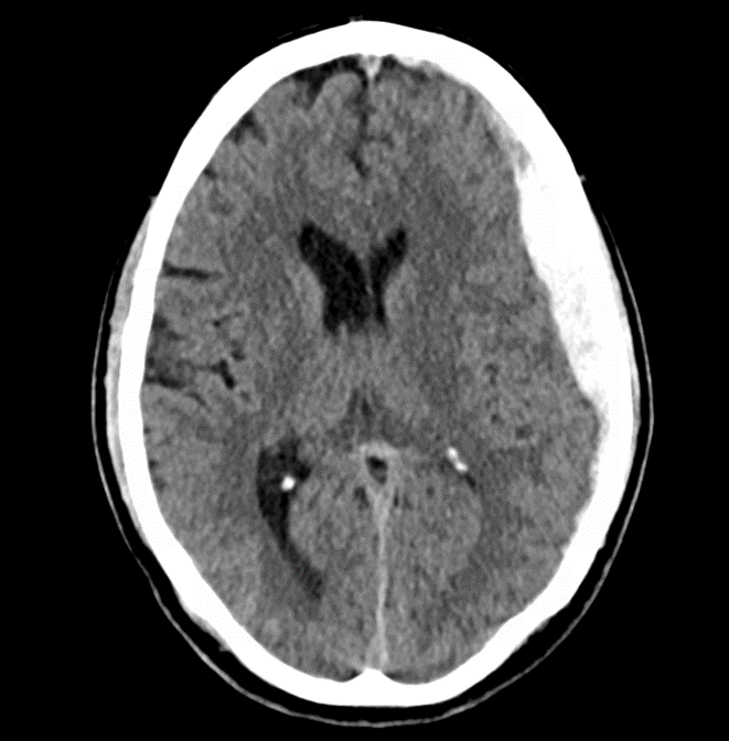
Answer: Subdural Hematoma
The patient’s anticoagulation was reversed with andexanet alfa. He additionally was provided with one unit of platelets and Keppra for seizure prophylaxis. Ultimately, the patient underwent embolization of the middle meningeal artery with significant clinical improvement of symptoms and was able to be discharged several days later.
Subdural Hematoma Pearls:
- Crescent shaped hematoma on CT
- Elderly and alcoholic patients at much higher risk due to brain atrophy
- Most commonly caused due to acceleration-deceleration injuries with tearing of the bridging veins
Eliquis reversal pearls:
- Traditionally 4-factor prothrombin complex concentrate (4Factor-PCC) has been used
- Contains Factors II, VII, IX, and X
- More recently, andexanet alfa has been developed which functions by binding and inhibiting factor Xa inhibitors
References:
Cline, David, et al. “Head Trauma.” Tintinalli’s Emergency Medicine: A Comprehensive Study Guide, McGraw-Hill Education, New York, 2020.