Tuesday Advanced Cases & Procedure Pearls

Pediatric Chest Pain by Dr. Edward Guo

Case: An 18 year old female with a past medical history of anxiety, 2 weeks post-op from tonsillectomy presents for chest pain. She woke up today with severe, non-radiating central chest pain. Her daily medications include sertraline and oral contraceptives. Vitals are BP 118/86, HR 119, Temp 97.7F, RR 24, SpO2 96% on room air. On exam, she is uncomfortable appearing and tearful. Her oropharynx is clear without signs of erythema or drainage. She is tachycardic without murmurs and has clear lung sounds bilaterally. There are no signs of lower extremity swelling. EKG and point-of-care cardiac ultrasound are shown below:

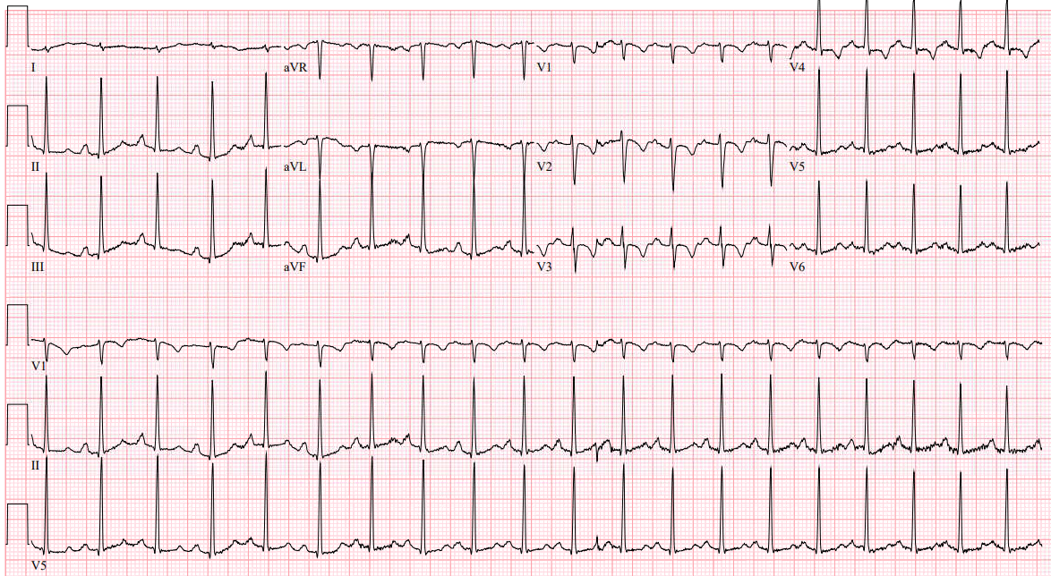
EKG interpretation: Sinus tachycardia with rightward axis and T wave inversions in anterior leads

POCUS interpretation: Normal LV ejection fraction. No pericardial effusion. Elevated RV to LV ratio. Dilated IVC with < 50% collapse with inspiration. RV free wall hypokinesis with apical hyperkinesis. Tricuspid annular plane systolic excursion 1.0 cm.

Case continued:  Due to high concern for pulmonary embolism, the patient is empirically started on heparin. CTA chest results with acute pulmonary embolism with right heart strain. High sensitivity troponin results markedly elevated at 306 mg/dL. Patient is admitted to the ICU with interventional cardiology consultation. Patient underwent pulmonary arteriogram which demonstrated elevated PA pressures without need for thrombectomy and was discharged in good condition on hospital day #4.

Pearls:

  • Infants and toddlers can have normal T wave inversions in the anterior leads known as “juvenile T waves” which should turn upright by age 7.
    • Any T wave inversion in these leads after they have flipped upright is abnormal.
  • While the S1Q3T3 EKG finding is classically associated with pulmonary embolism, more common EKG findings suggestive of PE include sinus tachycardia, rightward axis, incomplete right bundle branch block, and T wave inversions in the anterior leads.
  • Empirically start anticoagulation in patients with high concern for pulmonary embolism prior to definitive imaging.
  • Right ventricular free wall hypokinesis with apical hyperkinesis is known as McConnell’s sign and is pathognomonic for acute pulmonary embolism.
    • Other POCUS findings include an RV:LV ratio > 1 which is sensitive but not specific. A tricuspid annular plane systolic excursion (TAPSE) < 17 mm is a quantitative measure to diagnose right heart dysfunction on echocardiogram.

References:

Kline JA. Venous Thromboembolism Including Pulmonary Embolism. In: Tintinalli JE, Ma O, Yealy DM, Meckler GD, Stapczynski J, Cline DM, Thomas SH. eds. Tintinalli’s Emergency Medicine: A Comprehensive Study Guide, 9e. McGraw Hill; 2020.

Goodacre, S., & McLeod, K. (2002). ABC of clinical electrocardiography: Paediatric electrocardiography. BMJ (Clinical research ed.)324(7350), 1382–1385. https://doi.org/10.1136/bmj.324.7350.1382

Alerhand S, Hickey SM. Tricuspid Annular Plane Systolic Excursion (TAPSE) for Risk Stratification and Prognostication of Patients with Pulmonary Embolism. J Emerg Med. 2020;58(3):449-456. doi:10.1016/j.jemermed.2019.09.017

Tuesday Advanced Cases & Procedure Pearls

From the EMDaily Archives: BRASH Syndrome by Dr. Richard Byrne

Case: A 69 year old male with a past medical history of hypertension and type 2 diabetes arrives via EMS for chest pain and weakness. EMS found patient with a heart rate of 30, systolic BP 100, awake and and alert. Vitals upon arrival to the ED are BP 92/41, HR 42, Temp 97.7F, RR 20, SpO2 98% on room air. On exam, he is conversant with bradycardia and clear lung sounds. EKG is shown below:

EKG interpretation: sinus bradycardia with left bundle branch block pattern, Sgarbossa criteria negative

Differential diagnosis: acute cardiac ischemia, hyperkalemia, medication overdose (beta blockers, calcium channel blockers, digoxin, clonidine)

Management:

  • Transcutaneous pacer pads
  • 1 mg atropine given with no response
  • 500 mL IV fluid bolus improved SBP to 100
  • Cardiology consult with concerns for acute ischemia given chest pain and new left bundle branch block with bradycardia

Case continued: 40 minutes after arrival, patient suffered a brady-asystolic cardiac arrest. ACLS was started immediately and 1 mg epinephrine and 1 g calcium gluconate were administered. ROSC was achieved with 3 minutes, and patient became awake and alert. Critical lab values resulted with a potassium of 7.7 and creatinine of 3.7 (unknown baseline). The patient was admitted to the ICU on vasopressors, maintained adequate urine output, and did not require emergent hemodialysis. He was discharged in good condition two days later.

Diagnosis: BRASH Syndrome – Bradycardia, Renal failure, AV nodal blocking agents (beta blockers in this case), Shock, and Hyperkalemia

  • Check out this excellent summary of BRASH syndrome from the Journal of Emergency Medicine here.

Pearls:

  • Hyperkalemia is a common cause of bradycardia.
  • Unstable patients should be treated empirically with IV calcium while waiting for lab confirmation.
  • A VBG with lytes may provide a faster laboratory confirmation, though the specimen may have unrecognized hemolysis.
  • The combination of acute renal failure (sometimes from vomiting/diarrhea) in conjunction with AV nodal blocking agents may result in BRASH syndrome.
  • Acute treatment is aimed as for management of hyperkalemia and emergency hemodialysis may be required if patient is anuric or unresponsive to treatment.

References:

Farkas, Joshua et al. BRASH Syndrome: Bradycardia, Renal Failure, AV Blockade, Shock, and Hyperkalemia. Journal of Emergency Medicine 59 (2); 216-223.

Tuesday Advanced Cases & Procedure Pearls

Traumatic Oculocardiac Reflex by Dr. Tara Ferrante and Dr. Simon Sarkisian

Case: A 12-year-old male was hit in the right eye with a baseball just prior to arrival, now with headache, nausea, and vomiting, with swelling and pain around his right eye.  Patient was unable to look upward, and had pain with looking downward. When he presented to the Emergency Department, his initial heart rate was 44 BPM. Patient received emergent CT head which showed small volume retrobulbar hematoma, concern for entrapment of the right lateral rectus muscle, and acute mildly displaced right orbital floor fracture. Ophthalmology and OMFS were emergently consulted and recommended transfer for pediatric oculoplastic evaluation for retrobulbar hematoma, right orbital floor fracture, and high concern for entrapment.

Pearls:

  • A drop in heart rate by more than 20%, with associated nausea, syncope, or hypotension, after force is exerted on the extraocular muscles or globe of the eye, is known as the oculocardiac reflex. The bradycardia that develops is typically sinus bradycardia. In some cases, this reflex has been known to cause arrhythmias, asystole, and cardiac arrest.  This reflex can occur with facial trauma and nerve blocks, however, it is most commonly found during ophthalmologic surgery.
  • This reflex occurs with activation of the vagus nerve from nerve pathways originating from the stimulation of the opthalmic division of the trigeminal nerve.  The vagus nerve activation results in a diminished chronotropy, creating the response.

Treatment:

  • Removal of the cause, which in the setting of trauma, means consulting ophthalmology for an intervening procedure.
  • In the interim, patients can be stabilized using management for bradycardia, such as atropine, placing the patient on the pacing pads, and careful cardiac monitoring. 
  • If signs and symptoms are concerning for oculocardiac reflex in the setting of trauma, emergent CT and ophthalmology consult is recommended.  Evidence of ocular muscle entrapment, such as limited extra-ocular movements, diplopia, or proptosis, is an indication for emergent ophthalmology consult.

References:

1. Cavuoto, K, Barmettler, A, Prakalapakorn, G, Yen, M, Puente, M. Oculacardiac Reflex. EyeWiki from American Academiy of Opthalmology. March 30, 2023. https://eyewiki.aao.org/Oculocardiac_Reflex.

2. Dunville LM, Sood G, Kramer J. Oculocardiac Reflex. [Updated 2022 Sep 19]. In: StatPearls [Internet]. Treasure Island (FL): StatPearls Publishing; 2023 Jan-. Available from: https://www.ncbi.nlm.nih.gov/books/NBK499832/

3. Sires BS, Stanley RB Jr, Levine LM. Oculocardiac reflex caused by orbital floor trapdoor fracture: an indication for urgent repair. Arch Ophthalmol. 1998;116(7):955-956.

4. Walker RA, Adhikari S. Eye Emergencies. In: Tintinalli JE, Ma O, Yealy DM, Meckler GD, Stapczynski J, Cline DM, Thomas SH. eds. Tintinalli’s Emergency Medicine: A Comprehensive Study Guide, 9e. McGraw Hill; 2020. Accessed September 16, 2023. https://accessemergencymedicine-mhmedical-com.ezproxy.rowan.edu/content.aspx?bookid=2353&sectionid=222404436

Tuesday Advanced Cases & Procedure Pearls

Abdominal Cutaneous Nerve Entrapment Syndrome by Austin Chang, MS4

Case: A 29 year-old female with history of multiple ED visits presents for chronic abdominal pain. Previous workups all negative including CT imaging and EGD/colonoscopy. Vital signs are normal. Exam is notable for point tenderness to LLQ, not improved with pain medication. Repeat labs and CT negative for acute intra-abdominal abnormality. Further physical exam notable for positive Carnett’s test.

Definition: Abdominal Cutaneous Nerve Entrapment Syndrome (ACNES) is defined as entrapment of cutaneous branches of lower intercostal nerves at lateral border of rectus abdominis muscle. These branches make two 90 degree turns, travelling in fibrous sheaths along their course, and at these sharp angles the nerves are susceptible to entrapment.

Epidemiology: Prevalence of up to 2% in ED patients presenting with abdominal pain. Up to 30% incidence in patients with abdominal pain and negative prior workup. More common in young women.

Diagnosis: Carnett’s test. Localize pain to specific area, then patient tenses abdominal wall by performing a sit-up or raising legs. Test is considered positive if pain increases or remains the same (78% sensitive, 88% specific).

Treatment: First-line is combination injection at site of entrapment with corticosteroid and local anesthesia under ultrasound guidance.

References:

  1. Suleiman, S, Johnston, D. “The Abdominal Wall: An Overlooked Source of Pain” American Family Physician. August 2001.
  2. Kanakarajan, S., et al. “Chronic Abdominal Wall Pain and Ultrasound-Guided Abdominal Cutaneous Nerve Infiltration: A Case Series.” Pain Medicine, volume 12, Issue 3, 1 March 2011, Pages 382-386.
  3. Mol FMU, Maatman RC, De Joode LEGH, Van Eerten P, Scheltinga MR, Roumen R. Characteristics of 1116 Consecutive Patients Diagnosed With Anterior Cutaneous Nerve Entrapment Syndrome (ACNES). Ann Surg. 2021 Feb 1;273(2):373-378. doi: 10.1097/SLA.0000000000003224. PMID: 30817351.
Tuesday Advanced Cases & Procedure Pearls

Surgical Airway by Dr. Julia Shamis

A 58-year-old male with past medical history of neurofibromatosis status presents 4 days after emergent neuro IR embolization of a left occipital artery branches after feeling a pop in his surgical site followed by left-sided facial numbness radiating down to the left shoulder with word-finding difficulties. A large, expanding neck hematoma was noted on the left anterior neck. Patient was taken immediately for a CTA to attempt to identify the source of hemorrhage, and upon completion of CT imaging, the patient experienced cardiac arrest. Orotracheal intubation was unsuccessful due to the anatomic distortion from the expanding neck hematoma. Patient underwent emergent surgical cricothyroidotomy and achieved ROSC immediately thereafter.

Surgical Cricothyroidotomy PEARLS:

Indication: can’t intubate, can’t oxygenate, can’t ventilate

  • Generally after three attempts failed at orotracheal intubation and unable to maintain oxygenation
  • No oral access, masseter spasm, clenched teeth, trismus, structural deformities, laryngospasm, massive hemorrhage, mass effect/displacement of trachea, airway swelling, facial trauma, foreign bodies that cannot be removed from airway safely, no viable connection between upper and lower airway

Equipment: chlorhexidine or povidone iodine solution, 11 blade scalpel, bougie, ET tube

Procedure: The “knife, finger, bougie” technique

  • Palpate the cricothyroid membrane located inferior to the laryngeal prominence (i.e. Adam’s apple)
  • Stabilize the larynx using your thumb and middle finger while palpating the membrane with your index finger
  • Make a vertical incision along the cricothyroid membrane from the thyroid cartilage to the bottom of the cricoid cartilage. Palpate again with the index finger to confirm the cricothyroid membrane. Make a horizontal stab incision through the cricothyroid memrane and extend the incision 1 cm laterally
  • Remove the scalpel and insert the index finger into the trachea. Use your finger as a guide to pass the bougie through the opening. Continue insertion of the bougie until it “hangs up” in the lower pulmonary tract
  • Pass the tracheostomy tube or ET tube over the bougie to secure the airway.

References:

Boland C, Nasr NF, Voronov GG. Cricothyroidotomy. In: Reichman EF. eds. Reichman’s Emergency Medicine Procedures, 3e. McGraw Hill; 2018.

Walls R, Murphy M. Manual of Emergency Airway Management. 4th ed. Philadelphia, PA: Lippincott Williams; 2012.

Milner S, Bennett J. Emergency cricothyrotomy. The Journal of Laryngology & Otology. 1991;105(11):883-885.

Holmes J, Panacek E, Sakles J, Brofeldt B. Comparison of 2 Cricothyrotomy Techniques: Standard Method Versus Rapid 4-Step Technique. Annals of Emergency Medicine. 1998;32(4):442-446.

Tuesday Advanced Cases & Procedure Pearls

Euglycemic DKA by Dr. Sarah Perelman

A 45 year old male with a history of alcohol use disorder, diabetes on insulin, glipizide, and empagiflozin presents with chest pain and shortness of breath. 3 days ago, he drank 20 beers and then started vomiting. Last night, he developed chest pain and headache. Today, he has been feeling short of breath.

Vital signs: BP 126/70, HR 90, Temp 98.7F, RR 35, SpO2 99%. EKG is normal. Accucheck 182.

On exam, he is generally uncomfortable appearing and tachypneic with increased work of breathing. He has dry mucous membranes and clear lung sounds bilaterally. There is mild tenderness to palpation over the epigastric area.

Labs are notable for:

  • VBG: pH 7.08, pCO2 20, HCO3 9
  • BMP: Na 121, BUN 23, creatinine 1.12
  • Ethanol, salicylates, and acetaminophen levels undetectable
  • Lactate 1.3
  • Beta-hydroxybutyrate 9.8
  • Urine: 4+ ketones, normal specific gravity

Differential diagnosis includes: Euglycemic DKA, Alcoholic Ketoacidosis, Starvation Ketosis, and Toxic Alcohol Ingestion

Management:

  • Started with 1L NS bolus  
  • Insulin infusion @ 0.1 u/kg/hr 
  • Thiamine, folate supplementation 
  • GMAWs protocol for expected alcohol withdrawal 
  • Critical Care consultation

Pearls

  • Symptoms of acidosis: nausea, vomiting, headache, abdominal pain, generalized weakness, Kussmaul respirations (tachypnea with belly breathing and clear lungs)
  • Differential diagnosis for anion gap metabolic acidosis: uremia (high BUN/creat), lactic acidosis (sepsis/shock), ketoacidosis (DKA vs. alcoholic vs starvation), ingestion (salicylate vs. acetaminophen)
  • Euglycemic DKA: a rare disorder in which glucose level is relatively normal (<250) but ketoacidosis develops
  • Consider eDKA in pregnancy, type 1 diabetes, alcohol abuse, liver failure, starvation, but most notably in patients taking SGLT2 inhibitors (-“flozin”)
    • Treatment: D5NS + insulin, replete K if needed 
  • Euglycemic DKA and alcoholic ketoacidosis can be very difficult to distinguish, as alcohol use and poor PO intake can precipitate euglycemic DKA. In anyone with diabetes presenting like the case above who is on an SGLT2 inhibitor and impaired liver function, have a low threshold to start insulin to help drive the glucose into cells once glucose >180.

References:

Gabor, KD., Cline, DM. “Acid-Base Disorders.” Tintinalli’s Emergency Medicine a Comprehensive Study Guide, 9th Edition.” (73-78).

Howard RD, Bokhari SRA. Alcoholic Ketoacidosis. [Updated 2021 Dec 12]. StatPearls. Treasure Island (FL): StatPearls Publishing; 2022 Jan-. Available from: https://www.ncbi.nlm.nih.gov/books/NBK430922/

Mehta, A., Emmett M. “Fasting Ketosis and Alcoholic Ketoacidosis.” UpToDate. October, 2020.

Nyce, A. Byrne, R., Lubkin, C. Chansky, M. “Diabetic Ketoacidosis.” Tintinalli’s Emergency Medicine a Comprehensive Study Guide, 9th Edition.” (1433-1441).

Tuesday Advanced Cases & Procedure Pearls

Metformin Overdose by Dr. Eugene Marrone

51 y/o male with a PMH of DM presented with an out of hospital cardiac arrest after a suicide attempt, found to have a lactate of 26.9, bicarb of 3, and pH <6.8. After collateral from the family, it was found that he overdosed on metformin. 

METFORMIN OD:

Precise amount of metformin required to do this is unclear, but seems to be high (e.g., >20 grams). The main effect of metformin is inhibition of the mitochondrial transport chain complex-I, which essentially poisons the mitochondria.

Presentation

  • Vitals: The following abnormalities may be seen:
    • Hypothermia
    • Hypotension progressing to vasopressor-refractory shock can occur
  • GI symptoms often predominate: Nausea, vomiting, diarrhea, epigastric pain.
  • Delirium, decreased consciousness.

Management:

  • Supportive care, can consider bicarb 
  • Hemodialysis! Main indications:
    • Lactate >15-20 mM
    • pH <7.0-7.1
    • Failure to improve despite standard supportive measures

References:

Calello DP, Liu KD, Wiegand TJ, Roberts DM, Lavergne V, Gosselin S, Hoffman RS, Nolin TD, Ghannoum M; Extracorporeal Treatments in Poisoning Workgroup. Extracorporeal Treatment for Metformin Poisoning: Systematic Review and Recommendations From the Extracorporeal Treatments in Poisoning Workgroup. Crit Care Med. 2015 Aug;43(8):1716-30. doi: 10.1097/CCM.0000000000001002 [PubMed]

Wang GS, Hoyte C. Review of Biguanide (Metformin) Toxicity. J Intensive Care Med. 2019 Nov-Dec;34(11-12):863-876. doi: 10.1177/0885066618793385 [PubMed]

https://www.extrip-workgroup.org/metformin

Tuesday Advanced Cases & Procedure Pearls

From the EMDaily Archives: Refractory Trigeminal Neuralgia by Dr. Richard Byrne

A 34 year old female with a history of trigeminal neuralgia presented with a chief complaint of 5 days of severe, worsening paroxysms of pain in the left trigeminal nerve distribution. The pain was refractory to carbamazepine and gabapentin. Neurology was consulted and an unconventional therapy was recommended: 

  • Trigeminal neuralgia is a chronic facial pain syndrome characterized by paroxysms of severe, lancinating pain in the trigeminal nerve distribution: usually the maxillary or mandibular branches.
    • The pain is typically unbearable, having been described as “the worst pain a human can endure.” Before effective pharmacotherapy, patients would often resort to suicide.
  • First line therapy is carbamazepine at starting doses of 200 mg/day, titrated up as high as 1200 mg/day in divided doses.
  • Second line therapy is either gabapentin or lamotrigineBaclofen has been used as an add-on medication in refractory cases.
  • Severe pain exacerbations will often prompt patients to seek care in the ED and require opiates for acute relief.
  • Several case reports and case series have suggested intravenous phenytoin or fosphenytoin for abortive therapy, usually reporting complete or near complete relief of pain after infusion.
  • The patient received 1,000 mg of phenytoin over a 1.5 hour infusion. Reported pain decreased from 9/10 to 1/10 nearly immediately. She was discharged on oral oxcarbamazepine and baclofen with neurology follow-up.
  • Conclusion: Consider intravenous phenytoin or fosphenytoin for acute pain crises in trigeminal neuralgia patients.
    • Remember to infuse phenytoin no faster than 25-50 mg/min to avoid hypotension and bradycardia.

References
1. Cheshire, William. Fosphenytoin: An Intravenous Option for the Management of Acute Trigeminal Neuralgia Crisis.  Journal of Pain and Symptom Management 2001; 21(6): 506-510.

Tuesday Advanced Cases & Procedure Pearls

Advanced Cases: Poor Appetite with Dr. Julia Shamis

A 76 year old female with a history of advanced dementia is brought to the ED due to family concerns of poor PO intake for the last 7 days. Vitals are notable for a HR 109 and BP 70/40. On exam, she is cachectic, non-verbal, and somnolent. Point of care cardiac ultrasound demonstrates the following:

Diagnosis: Pericardial Tamponade

Teaching Pearls:

  • Presenting signs/symptoms
    • Most common symptoms: dyspnea, chest pain, syncope, and altered mental status
    • Exam may demonstrate: JVD, tachycardia, hypotension, muffled heart sounds, narrowed pulse pressure, and tachypnea
  • Point of care ultrasound findings:
    • Effusions can be visualized in all four traditional cardiac views
    • Tamponade occurs when the pericardial pressure > intracardiac pressures, resulting in RV collapse in diastole (most specific finding for tamponade)
    • Right atrial collapse in early diastole can be first sign of cardiac tamponade (more sensitive but not as specific)
    • LV ejection fraction is often high to compensate for decreased filling volume

Treatment:

  • IV fluid resuscitation
  • In hemodynamically stable patients, treat with pericardiocentesis under fluoroscopy with interventional cardiology or to the OR for pericardial window with cardiothoracic surgery
  • If hemodynamically unstable, bedside pericardiocentesis should be performed, ideally under direct ultrasound guidance

References

Franz, Taylor. “Pericardial Effusion and Cardiac Tamponade: Pearls and Pitfalls.” EmDOCs.net – Emergency Medicine Education, 4 Apr. 2022, www.emdocs.net/pericardial-effusion-and-cardiac-tamponade-pearls-and-pitfalls/. Accessed 12 July 2023.

Stashko E, Meer JM. Cardiac Tamponade. [Updated 2022 Aug 8]. In: StatPearls [Internet]. Treasure Island (FL): StatPearls Publishing; 2023 Jan-. Available from: https://www.ncbi.nlm.nih.gov/books/NBK431090/

Farkas, Josh. “Pericardial Tamponade.” EMCrit Project, 10 Nov. 2021, emcrit.org/ibcc/tamponade/.

Alerhand S, Adrian RJ, Long B, Avila J. Pericardial tamponade: A comprehensive emergency medicine and echocardiography review. Am J Emerg Med. 2022;58:159-174. doi:10.1016/j.ajem.2022.05.001