Wednesday Image Review

What’s the Diagnosis? By Jake Barr, MS3

The patient is a 40-year-old male with no past medical history who presents with concerns of a rash on his hands. He states that the lesions appeared two days ago, but had a fever, muscle aches, and pruritis two days before that. He does not have a history of hives or contact dermatitis. He does not take any medications, but states he recently developed “cold sores.” His temperature is 100.6oF, but his other vitals are within normal limits. The cutaneous rash is demonstrated below. When looking in his mouth, blistering lesions are also present. What the diagnosis and management?

Answer: Erythema Multiforme

  • Erythema multiforme is the result of a T-cell mediated hypersensitivity reaction resulting in a characteristic pruritic, targetoid papules, with a hazy-center, and surrounding erythematous rings.
  • 90% of cases are associated with infectious etiologies, with HSV-1 being most common in adults and Mycoplasma pneumonia in children.
    • Remaining 10% are due to drugs (NSAIDs, antiepileptics, antibiotics), malignancy, and autoimmune diseases. Their mechanism of rash formation is unknown.
  • There are two specific subtypes:
    • Erythema multiforme minor: rash without mucosal involvement or constitutional symptoms
    • Erythema multiforme major: rash with mucous membrane involvement and constitutional symptoms (fever, malaise, myalgias etc.)
  • Diagnosis is often clinical, but immunofluorescence can be helpful if uncertain.
    • PCR testing for both HSV and Mycoplasma pneumoniae should be done if suspected.
  • Treatment is mostly symptomatic, and the rash is usually self-limited.
    • Antihistamines are useful for pruritis.
    • Systemic steroids maybe be used, but the impact on long-term outcomes and symptom duration is unclear.
    • Acyclovir may be used to prevent recurrent HSV infections.
    • Macrolides may be used in Mycoplasma pneumoniae is suspected.

References:

  1. Baluzy Matthew, Karaze Tallib. Maculopapular Rashes. In: Mattu A and Swadron S, ed. CorePendium. Burbank, CA: CorePendium, LLC. https://www.emrap.org/corependium/chapter/recvFPlc0BmXxYuzp/Maculopapular-Rashes#h.til8vwjxmfh6. Updated June 21, 2023. Accessed January 24, 2024.
  2. Sokumbi O, Wetter DA. Clinical features, diagnosis, and treatment of erythema multiforme: a review for the practicing dermatologist. Int J Dermatol. 2012;51(8):889-902. doi:10.1111/j.1365-4632.2011.05348.x
  3. Trayes KP, Love G, Studdiford JS. Erythema Multiforme: Recognition and Management. Am Fam Physician. 2019;100(2):82-88.
  4. J. Brady W, Pandit A, R. Sochor M. Generalized Skin Disorders. In: Tintinalli JE, Ma O, Yealy DM, Meckler GD, Stapczynski J, Cline DM, Thomas SH. eds. Tintinalli’s Emergency Medicine: A Comprehensive Study Guide, 9e. McGraw-Hill Education; 2020. Accessed January 24, 2024. https://accessemergencymedicine.mhmedical.com/content.aspx?bookid=2353&sectionid=221180403
  5. DermNet. https://dermnetnz.org/topics/erythema-multiforme-images
Tuesday Advanced Cases & Procedure Pearls

Critical Cases – TCA Overdose!

by Daniel Petrosky M.D.

HPI

  • 31 y.o. female presents with acute change in mental status
  • Family found unresponsive 
  • EMS trialed one dose of naloxone without effect

PMHX

  • Multiple sclerosis, chronic pain, opioid use disorder, generalized anxiety disorder, and major depressive disorder,

Physical Exam

  • Markedly dry mucous membranes and cracked, dry lips 
  • Pt lethargic, localizes pain, mumbles, and does not follow commands
  • Afebrile

Work-up

  • ECG shows prolonged QT otherwise unremarkable
  • BMP, CBC, LFTs, acetaminophen, salicylate , UA all WNL
  • UDS positive for TCH, benzos, and amphetamines 
  • Bladder scan and subsequent bladder catheterization reveal over 1 L clear urine

Case Conclusion

  • Several hours later the pt was able to state that she overdosed on her amitriptyline and wrote a suicide note
  • Toxicology consulted did not recommend any acute interventions
  •  Psychiatry consulted for suicide attempt. 

TCA Overdose Pearls

  • Toxicity can vary in presentation and thorough review of medications as well as collateral from family can be very important
  • TCA overdose can be tricky as it can affect multiple organ systems and present with anti-cholinergic properties (see below), ECG changes such as QTc prolongation, QRS prolongation, and a “terminal r wave” in lead aVR , and seizures.
  • Symptoms typically occur 6 hours after ingestion and can be worse with con-ingestion of sedatives
  • Those with ECG changes should be monitored for 36-48 hrs
  • Treatment is aimed at overcoming cardiac sodium channel blockade with sodium bicarbonate or hypertonic saline, and is reserved for those patients with ECG changes
Classic Symptom DescriptionPhysical Exam Manifestation
“Mad as a Hatter”Acute encephalopathy
“Red as a Beet”Erythroderma (in fair skinned patients)
“Blind as a Bat”Dilated and unresponsive pupils
“Dry as a Bone”Dry, cracked mucous membranes, no sweating
“Tachy as a Leisure Suit”Sinus tachycardia
“Hot as Hell”Hyperthermia

References:

In: Tintinalli JE, Stapczynski J, Ma O, Yealy DM, Meckler GD, Cline DM. eds. Tintinalli’s Emergency Medicine: A Comprehensive Study Guide, 8e. McGraw Hill; 2016, 1194-1199.

Monday Back to Basics & Pharmacology

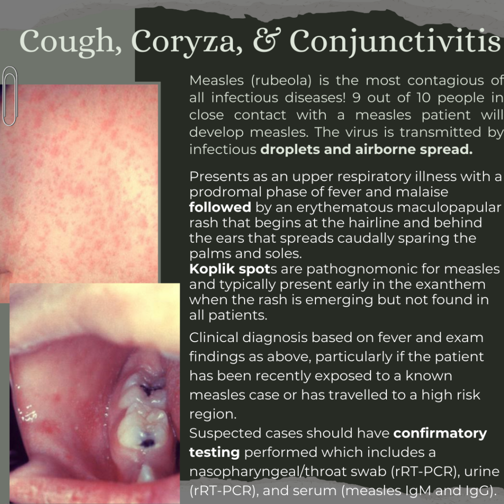
Measles Part 1: Identification with Drs. Edward Guo and Simon Sarkisian

While most cases of measles are mild and will self-resolve, the high infectivity of the virus is a public health hazard due to rare complications of the disease that can cause long-term morbidity and mortality. Particularly high risk populations include unvaccinated individuals, children < 5 years, adults > 20, pregnant women, and immunocompromised patients.  

Look forward to part 2 for more details on measles management, treatment, and complications! 

References: 

https://www.nj.gov/health/cd/topics/measles.shtml

https://www.cdc.gov/measles/hcp/index.html

Takhar SS, Moran GJ. Serious Viral Infections. In: Tintinalli JE, Ma O, Yealy DM, Meckler GD, Stapczynski J, Cline DM, Thomas SH. eds. Tintinalli’s Emergency Medicine: A Comprehensive Study Guide, 9e. McGraw-Hill Education; 2020. 

Nguyen M, Dunn AL. Rashes in Infants and Children. In: Tintinalli JE, Ma O, Yealy DM, Meckler GD, Stapczynski J, Cline DM, Thomas SH. eds. Tintinalli’s Emergency Medicine: A Comprehensive Study Guide, 9e. McGraw-Hill Education; 2020. 

Friday Board Review

Board Review by Dr. Hilbmann (Edited by Dr. Parikh)

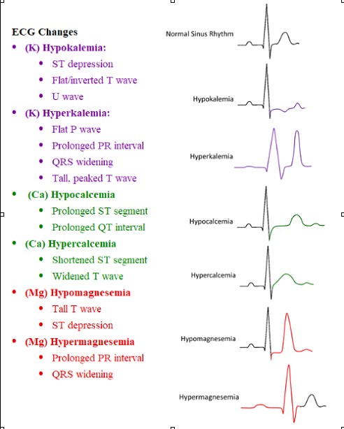
A 22 year old female G1P0 with no pmhx who presents to the Emergency Department with nausea and vomiting. The patient is 11 weeks pregnant and has experienced nausea and occasional vomiting throughout her pregnancy but for the past three days she has been progressively worse and has not been able to keep any food or liquids down without vomiting. You are given her ECG, what is a finding you are looking for due to her recent presentation?


A. Hyperacute T waves
B. Shortened PR Interval
C. Prolonged QT interval
D. ST-Elevation in aVR

Answer: C.
Due to this patient’s increased vomiting, most likely due to her pregnancy, it is possible that she may be experiencing electrolyte imbalances secondary to the loss of gastrointestinal contents. Most commonly hypokalemia, hypocalcemia, and hypomagnesemia can result from continuous vomiting. In addition to blood tests, which take time to result, the levels of these three electrolytes can be evaluated with ECG as well. Physicians should evaluate for increased p waves, prolonged PR interval, ST depression, u waves, and a prolonged QT interview.

References:
Mitchell SJ, Cox P. ECG changes in hyperemesis gravidarum. BMJ Case Rep. 2017;2017(bcr2016217158) doi: 10.1136/bcr-2016-217158.
Popa SL, Barsan M, Caziuc A, Pop C, Muresan L, Popa LC, Perju-Dumbrava L. Life-threatening complications of hyperemesis gravidarum. Exp Ther Med. 2021 Jun;21(6):642. doi: 10.3892/etm.2021.10074. Epub 2021 Apr 16. PMID: 33968173; PMCID: PMC8097228.
Image per @medicalce via Twitter

Wednesday Image Review

From the Archives: What’s the Diagnosis? By Dr. Alyse Volino

A 24 year old female with a history of gallstones presents to the ED with 2 hours of severe, aching right upper quadrant abdominal pain that woke her from sleep associated with nausea and vomiting. Patient has had similar episodes of pain in the past, often after eating fatty foods. On exam, she is tender in RUQ of the abdomen and is actively vomiting. A right upper quadrant abdominal ultrasound is performed and shown below. What’s the diagnosis?

Answer: Symptomatic Cholelithiasis

  • Characterized by episodes of RUQ pain that are brought on by obstructing gallstone and relieved when gallstone moves from that position
  • Can progress to cholecystitis if gallstone remains in obstructing position and gallbladder subsequently becomes inflamed or infected.
  • Differential: cholecystitis, choledocholithiasis, cholangitis.
  • If simple symptomatic cholelithiasis (no signs of infection or other biliary obstruction) and pain is controlled, patient can often be discharged from ED with outpatient surgical follow-up.

Symptomatic Cholelithiasis vs Cholecystitis on Ultrasound

Cholecystitis may have the following:

  1. Gallstones present in gallbadder
  2. Sonographic Murphy’s sign
    • Maximal tenderness over the most anterior portion of GB as defined with US imaging
  3. Wall thickness of gallbladder over 3 mm
    • Be sure to measure GB wall thickness at anterior aspect to avoid false positive increased secondary to posterior acoustic enhancement!
  4. Pericholecystic fluid

These findings should be used in conjunction with lab results to help identify diagnosis on spectrum of biliary disease.

References:

Tintinalli’s Emergency Medicine: A Comprehensive Study Guide, 8th Edition. Ch 79: Pancreatitis and Cholecystitis. Judith E. Tintinalli

Soni, Nilam, et. al. Point of Care Ultrasound, 2nd Edition. Ch 27: Gallbladder 

Monday Back to Basics & Pharmacology

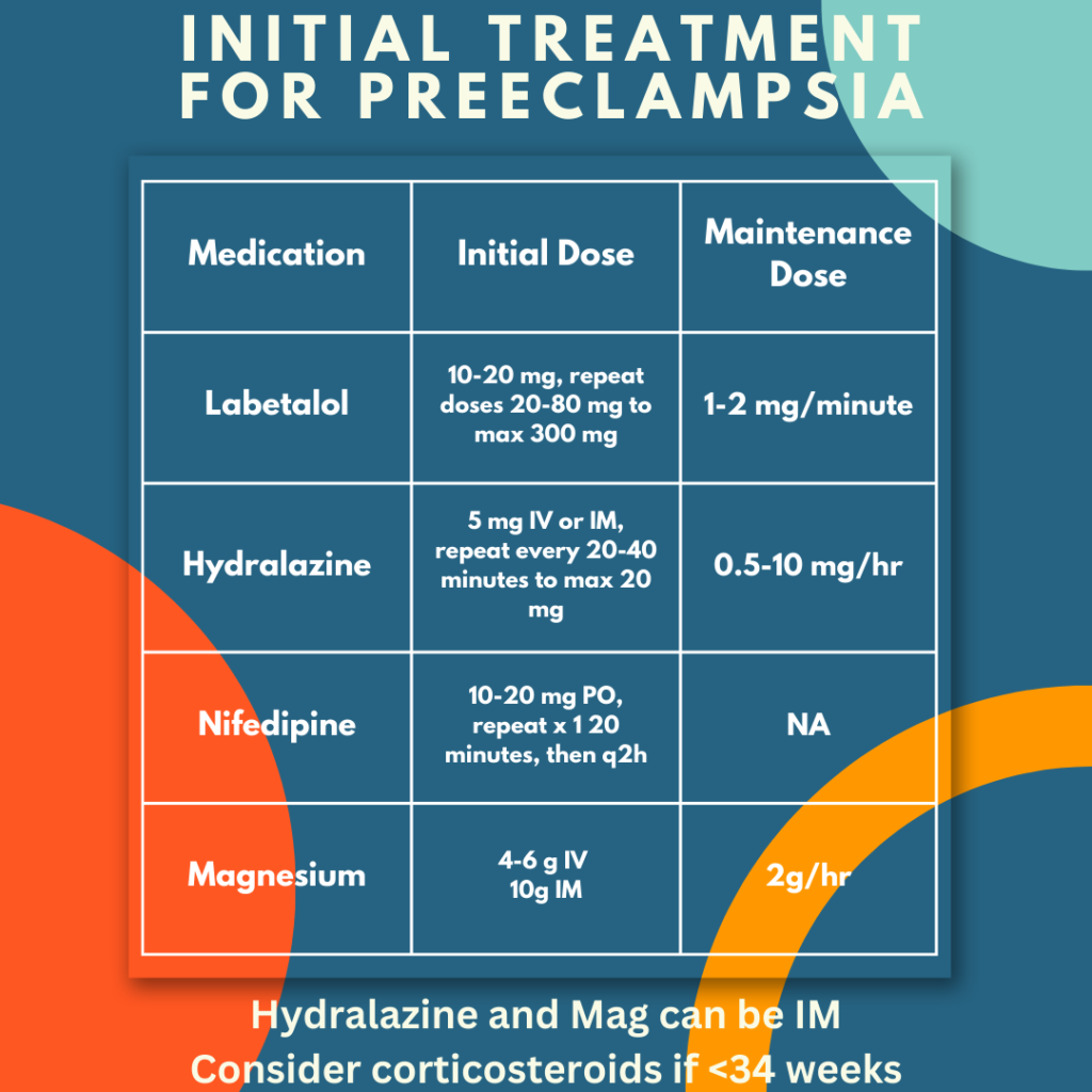
Preeclampsia Management with Dr. Erica Westlake

Use these medications for aggressive blood pressure control

Load patients with magnesium early

Ultimate treatment is delivery – involve OB/NICU teams early, transfer patients if these teams are not available

Referneces:

Friday Board Review

Board Review by Alex Hilbmann

Vital signs:

T: 37⁰C HR: 71 bpm O2 Sat: 100% BP: 112/92

A 29 year old female reports to the emergency department for abnormal scant vaginal bleeding. Initially, she believed that the bleeding was an early menstrual period but it has now persisted longer than her usual menses with less volume. She denies any other complaints, including pelvic pain, fevers, or vaginal discharge. Patient has attended OB/GYN appointments yearly and denies any previous history of sexually transmitted infections or abnormal pap smears. Pelvic exam reveals scant bleeding from the cervical os with no adnexal or cervical tenderness. No masses are appreciated upon palpation of bilateral adnexa. Transvaginal ultrasound reveals no intrauterine pregnancy or adnexal abnormalities. Point of care urine pregnancy test is positive. Quantitative beta-hcg results at 542 miU/mL. Vital Signs listed above. What is the next best step in management of this patient?

  1. Consultation to OB/GYN for concern of ectopic pregnancy
  2. Official transvaginal ultrasound read by Radiology
  3. Administration of methotrexate and discharge home
  4. Discharge home with 2 day OB/GYN follow up

Answer: D.

Our patient is currently experiencing scant vaginal bleeding with no findings on transvaginal ultrasound, a positive pregnancy test, and a beta hcg below 1500 miU/mL.  1500 miU/mL is what is known as the “discriminatory zone” for transvaginal ultrasound (6,000 miU/mL for transabdominal). This discriminatory zone is a level of beta-hcg which an intrauterine pregnancy (IUP) would be expected to be seen on ultrasound. When beta-hcg is above the discriminatory zone, and therefore an IUP should be visible on ultrasound, the absence of an IUP on ultrasound suggests ectopic pregnancy until proven otherwise. With this patient however, we are below the discriminatory zone and therefore the absence of IUP (with no other findings suggesting ectopic pregnancy) does not make ectopic pregnancy a more likely diagnosis than pregnancy with implantation bleeding at this time. The beta hcg of an IUP should double by two days, whereas an ectopic pregnancy would decrease less than twofold. This can be assessed by the patient’s OB/GYN at her follow up, and given the patient’s stable condition she can be discharged home.

Tintinalli, J., Ma, O., Yealy, D., Meckler, G., Cline, D., Thomas, S. and Stapczynski, J., 2020. Tintinalli’s emergency medicine. 9th ed. [New York]: McGraw-Hill Education, pp.615-620.

Image:

Fadial, T., 2018. Differential Diagnosis of Ultrasound in Ectopic Pregnancy. [online] Differential Diagnosis of. Available at: <https://ddxof.com/ultrasound-in-ectopic-pregnancy/?sf_action=get_data&sf_data=all&_sf_s=ectopic> [Accessed 2 October 2022].

Wednesday Image Review

What’s the Diagnosis? By Dr. Edward Guo

A 2 year old female with no past medical history presents for a nasal foreign body. The patient’s mother states that the child approached her earlier this evening while pointing at her nose. When the mother looked into the child’s right nostril, she noticed something metal. Vital signs are normal for age. On exam, the patient is in no acute distress with mucus coming from the right nostril and a silver metallic object lodged in the nare. A skull x-ray is obtained and shown below. The area of interest is included and zoomed in. What’s the diagnosis and what’s the appropriate management?

Answer: Nasal button battery – emergent removal in ED

  • Button batteries are distinguished on plain films most commonly by the “double ring sign” in AP view which is best shown in the lateral film above. Lateral views of a button battery may also show a step-off which is also evident in the AP x-ray.
  • Management of a nasal button battery differs from many other foreign bodies due to the risk of rapid necrosis and septal perforation in as little as 7 hours.
  • ED methods for removal of nasal foreign bodies include the “parent kiss” technique or bag valve mask to expel the foreign body, forcep or suction catheter removal, or use of a foley balloon.
  • In this case, the object was ultimately removed by ENT with procedural sedation in the ED.

References:

Cohen JS, Agrawal D. Nose and Sinus Disorders in Infants and Children. In: Tintinalli JE, Ma O, Yealy DM, Meckler GD, Stapczynski J, Cline DM, Thomas SH. eds. Tintinalli’s Emergency Medicine: A Comprehensive Study Guide, 9e.

McGraw Hill; 2020.Loh  WS, Leong  J, Tan  HK: Hazardous foreign bodies: complications and management of button batteries in the nose. Ann Otol Rhinol Laryngol 112: 379, 2003. [PubMed: 12731636]