

")


Alyssa Exarchakis, MS4


Alyssa Exarchakis, MS4

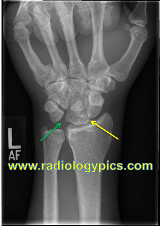
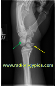
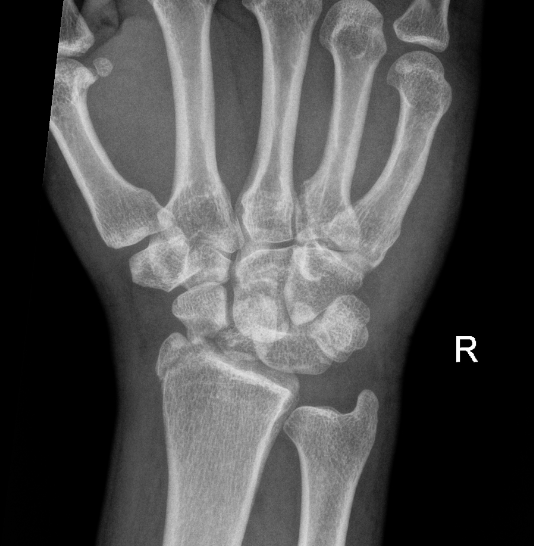
A 38 year old male presents to the ED with a chief complaint of right wrist pain that began after a fall off a motorcycle the day prior. On exam, the patient is noted to have tenderness along his right distal radius, snuffbox tenderness, wrist swelling, and is unable to flex/extend the wrist. Normal pulses and sensation is present.
Wrist X-rays demonstrate the following:
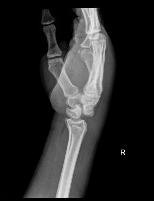
What is the diagnosis?
Perilunate dislocation, scaphoid fracture, radial styloid fracture
Perilunate Dislocation:
References:
Cheffers M. Wrist Reduction Techniques. In: Johnson W, Nordt S, Mattu A and Swadron S, eds. CorePendium. Burbank, CA: CorePendium, LLC. https://www.emrap.org/corependium/chapter/recoPqKOBgkCSHesR/Wrist-Reduction-Techniques#h.493xci6kkby6. Updated December 21, 2022. Accessed August 15, 2024.
Mark Karadsheh. “Lunate Dislocation (Perilunate Dissociation).” Orthobullets, 5 Nov. 2022, www.orthobullets.com/hand/6045/lunate-dislocation-perilunate-dissociation. “Solution to Unknown Case #30 – Perilunate Dislocation.” RADIOLOGYPICS.COM, 6 Jan. 2014, radiologypics.com/2013/03/28/perilunate-dislocation/.

A 5-year-old boy presents to the emergency department with a two-day history of fever, sore throat, and difficulty swallowing. On examination, he has multiple small, grayish-white papulovesicular lesions on the soft palate, uvula, and tonsillar pillars. He is otherwise alert and well-hydrated with stable vital signs. Which of the following is the most likely diagnosis?
A) Peritonsillar abscess
B) Herpangina
C) Hand-foot-and-mouth disease
D) Infectious mononucleosis
E) Scarlet fever
Explanation:
Herpangina is characterized by the sudden onset of fever, sore throat, and dysphagia, accompanied by small, vesicular lesions on the posterior oropharynx, typically involving the soft palate, uvula, and tonsillar pillars. The lesions are grayish-white and may be surrounded by erythema. It is caused by Coxsackievirus group A, primarily affecting young children. Treatment is supportive, focusing on pain management and hydration. Peritonsillar abscess (choice A) presents with severe throat pain, trismus, and unilateral tonsillar swelling. Hand-foot-and-mouth disease (choice C) manifests with oral ulcers and vesicles on the hands and feet. Infectious mononucleosis (choice D) presents with fever, sore throat, lymphadenopathy, and atypical lymphocytosis. Scarlet fever (choice E) presents with a sandpaper-like rash and strawberry tongue, secondary to group A Streptococcus infection.
Therefore, the correct answer is B) Herpangina

References: Tintinalli’s Emergency Medicine Manual, 9th Edition

Case: 38 y/o female with a history of bilateral renal stones and recent lithotripsy for renal stone who presented with left flank pain and nausea. Symptoms were consistent with previous renal colic symptoms. Following lithotripsy, the patient had resolution of symptoms before flank pain and nausea returned 3 days ago. Vitals BP 142/97, Pulse 93, Temp 98.1 °F (36.7 °C) (Oral), Resp 18, SpO2 99%. The physical exam demonstrated left sided abdominal tenderness and left CVA tenderness. Bedside ultrasound findings below.
Answer: Ureteral stone at the left UPJ with mild-moderate left hydronephrosis
Ultrasound findings in nephrolithiasis
Hyperechoic foci with posterior acoustic shadowing
Hydronephrosis
Twinkle Sign
Ureteral jets

Conclusions:
Ultrasound can be used first-line for imaging to assess for renal stones, though may require follow-up imaging
Ultrasound may prevent repeated radiation exposure with CT in patients with known renal stones
Bedside US can allow for rapid diagnosis and treatment as well as faster discharge when assessing for renal stones
References:
1. Coursey CA, Casalino DD, Remer EM, Arellano RS, Bishoff JT, Dighe M, et al. ACR Appropriateness Criteria® acute onset flank pain–suspicion of stone disease. Ultrasound Q. 2012 Sep. 28 (3):227-33
2. Dillman JR, Kappil M, Weadock WJ, Rubin JM, Platt JF, DiPietro MA, Bude RO. Sonographic twinkling artifact for renal calculus detection: correlation with CT. Radiology. 2011 Jun;259(3):911-6. doi: 10.1148/radiol.11102128. Epub 2011 Apr 1. PMID: 21460031.
4. Gliga, M. L., Chirila, C. N., Podeanu, D. M., Imola, T., Voicu, S. L., Gliga, M. G., & Gliga, P. M. (2017). Twinkle, twinkle little stone: an artifact improves the ultrasound performance! Medical Ultrasonography, 19(3), 272-275. https://doi.org/10.11152/mu-984
3. Ongun S, Teken A, Yılmaz O, Süleyman S. Can Ureteral Jet Flow Measurement Predict Spontaneous Passage of Distal Ureteral Stones? Urol Int. 2018;101(2):156-160. doi: 10.1159/000490498. Epub 2018 Jun 27. PMID: 29949810.
4. Wong C, Teitge B, Ross M, Young P, Robertson HL, Lang E. The Accuracy and Prognostic Value of Point-of-care Ultrasound for Nephrolithiasis in the Emergency Department: A Systematic Review and Meta-analysis. Acad Emerg Med. 2018 Jun;25(6):684-698. doi: 10.1111/acem.13388. Epub 2018 Mar 25. PMID: 29427476.
5. Brisbane W, Bailey MR, Sorensen MD. An overview of kidney stone imaging techniques. Nat Rev Urol. 2016 Nov;13(11):654-662. doi: 10.1038/nrurol.2016.154. Epub 2016 Aug 31. PMID: 27578040; PMCID: PMC5443345.

References:


References:
Tintinalli’s Emergency Medicine: A Comprehensive Study Guide, 9e Tintinalli JE, Ma O, Yealy DM, Meckler GD, Stapczynski J, Cline DM, Thomas SH. Tintinalli J.E., & Ma O, & Yealy D.M., & Meckler G.D., & Stapczynski J, & Cline D.M., & Thomas S.H.(Eds.),Eds.
EMRap compendium
Konca C, Kahramaner Z, Bosnak M, Kocamaz H. Hemlock (Conium Maculatum) Poisoning In A Child. Turk J Emerg Med. 2016 Feb 26;14(1):34-6. doi: 10.5505/1304.7361.2013.23500. PMID: 27331163; PMCID: PMC4909876.