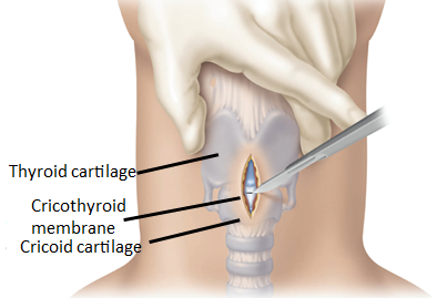
Friday Board Review

Board Review with Dr. Alex Hilbmann

A 28 year old female G4P2 at 8 weeks gestation presents to the Emergency Department after vomiting almost four times daily for the past week. She denies any recent fevers, abdominal pain, pelvic pain, or vaginal bleeding. Vital signs include: Temp 37.2 C, HR 105, BP 93/62, SpO2 100%. On exam, she is uncomfortable appearing with dry mucous membranes and intermittently dry heaving into an emesis bag. Blood serum results are pending. Urinalysis reveals 1+ ketones with elevated specific gravity. What is the next best step in management?

A. 0.9% normal saline

B. Prophylactic electrolyte repletion

C. 5% dextrose and 0.9% normal saline

D. Antiemetic and PO challenge

Answer: 5% dextrose and 0.9% normal saline

This pregnant patient is most likely experiencing hyperemesis gravidarum given her presentation of multiple episodes of vomiting, volume depletion, and ketonuria. The treatment for hyperemesis gravidarum includes 5% glucose in IV fluids, anti-emetic drugs, and correction of any electrolyte abnormalities. Nothing should be given by mouth until patient’s nausea is controlled, and although this patient will ultimately benefit from antiemetic administration with the hopes that she will tolerate PO, her signs of volume depletion and ketonuria suggest immediate treatment with 5% dextrose in 0.9% normal saline or lactated ringer solution.

Management of Hyperemesis Gravidarum
First line: pyridoxine (vitamin B6) – pregnancy drug class A
Add on: doxylamine – pregnancy drug class A
Adjuncts: ondansetron, metoclopramide – pregnancy drug class B
IV fluids with dextrose

References:

References: Tintinalli, J., Ma, O., Yealy, D., Meckler, G., Cline, D., Thomas, S. and Stapczynski, J., 2020. Tintinalli’s emergency medicine. 9th ed. [New York]: McGraw-Hill Education, pp.621-622.

Nausea and vomiting of pregnancy. ACOG Practice Bulletin No. 153. American College of Obstetricians and Gynecologists. Obstet Gynecol. 2015; 126(3):e12-24

Wednesday Image Review

What’s the Diagnosis? By Dr. Carlos Cevallos

A 55 year old male with a past medical history of colon cancer on chemotherapy presents with a chief complaint of right lower extremity pain/discoloration to his calf and thigh as well shortness of breath that has developed over the past 48 hours. A physical exam reveals dopplerable DP and PT pulses. The right calf and thigh is visualized as in the image below. What’s the diagnosis?

Answer: Phlegmasia Cerulea Dolens (PCD) – a near-total occlusion of the major deep venous system of an extremity as well as the majority of microvascular collateral veins of the extremity. 

PCD occurs on a spectrum with phlegmasia albans dolens (PAD): thrombosis of the deep venous system with patency of the collateral veins and venous gangrene: when there is complete obstruction of venous outflow with irreversible capillary involvement and muscle infarction. It is differentiated from PAD by a pale/white limb versus a dusky/cyanotic limb in PCD. 

Clinical Features: Triad of swelling, pain, cyanosis. Limb can develop firmness and there is a risk for arterial compromise and compartment syndrome. Thrombosis can extend into the IVC and it is often accompanied by pulmonary embolism with the incidence reported to vary from 12-40%.  

Diagnosis: Clinical history/exam in conjunction with imaging. Gold standard diagnosis is contrast venography, however often due to difficulty and length of time to attain this ultrasound venography is often preferred. CT-venogram is useful for visualization of extension of thrombus in the IVC. 

Management: Immediate elevation of affected extremity above the level of the heart to encourage return of circulation. Anticoagulation with unfractionated IV heparin bolus at 10-15 units/kg followed by an infusion titrated to an aPTT of 1.5-2 times the lab control value. Immediate vascular/interventional radiology consultation for possibly thrombectomy versus catheter-directed thrombolysis. If no service is available and transfer is unable to be arranged within 6 hours then consider systemic fibrinolytics if no contraindications are present. 

Case Continued: Duplex ultrasound, CT-venogram, and CTA Chest on our patient revealed DVT of the major deep veins of the right lower extremity that extended into the IVC as well as bilateral pulmonary embolisms. He was started on heparin and had a mechanical thrombectomy of the right iliofemoral/IVC DVT. He was able to be discharged on apixaban several days later. 

Resources:

Cline, D., Ma, O. J., Meckler, G. D., Stapczynski, J. S., Thomas, S. H., Tintinalli, J. E., Yealy, D. M., & Kline, J. A. (2020). Venous Thromboembolism Including Pulmonary Embolism. In Tintinalli’s emergency medicine: A comprehensive study guide (pp. 389–398). essay, McGraw-Hill Education. 

Gardnella, L., & Falk, J. (n.d.). Phlegmasia Alba and cerulea Dolens – StatPearls – NCBI Bookshelf. Phlegmasia Alba and Cerulea Dolens. https://www.ncbi.nlm.nih.gov/books/NBK563137/ 

Perkins, J. M., Magee, T. R., & Galland, R. B. (1996). Phlegmasia caerulea dolens and venous gangrene. British Journal of Surgery, 83(1), 19–23. https://doi.org/10.1002/bjs.1800830106 

Tuesday Advanced Cases & Procedure Pearls

Abdominal Cutaneous Nerve Entrapment Syndrome by Austin Chang, MS4

Case: A 29 year-old female with history of multiple ED visits presents for chronic abdominal pain. Previous workups all negative including CT imaging and EGD/colonoscopy. Vital signs are normal. Exam is notable for point tenderness to LLQ, not improved with pain medication. Repeat labs and CT negative for acute intra-abdominal abnormality. Further physical exam notable for positive Carnett’s test.

Definition: Abdominal Cutaneous Nerve Entrapment Syndrome (ACNES) is defined as entrapment of cutaneous branches of lower intercostal nerves at lateral border of rectus abdominis muscle. These branches make two 90 degree turns, travelling in fibrous sheaths along their course, and at these sharp angles the nerves are susceptible to entrapment.

Epidemiology: Prevalence of up to 2% in ED patients presenting with abdominal pain. Up to 30% incidence in patients with abdominal pain and negative prior workup. More common in young women.

Diagnosis: Carnett’s test. Localize pain to specific area, then patient tenses abdominal wall by performing a sit-up or raising legs. Test is considered positive if pain increases or remains the same (78% sensitive, 88% specific).

Treatment: First-line is combination injection at site of entrapment with corticosteroid and local anesthesia under ultrasound guidance.

References:

  1. Suleiman, S, Johnston, D. “The Abdominal Wall: An Overlooked Source of Pain” American Family Physician. August 2001.
  2. Kanakarajan, S., et al. “Chronic Abdominal Wall Pain and Ultrasound-Guided Abdominal Cutaneous Nerve Infiltration: A Case Series.” Pain Medicine, volume 12, Issue 3, 1 March 2011, Pages 382-386.
  3. Mol FMU, Maatman RC, De Joode LEGH, Van Eerten P, Scheltinga MR, Roumen R. Characteristics of 1116 Consecutive Patients Diagnosed With Anterior Cutaneous Nerve Entrapment Syndrome (ACNES). Ann Surg. 2021 Feb 1;273(2):373-378. doi: 10.1097/SLA.0000000000003224. PMID: 30817351.
Monday Back to Basics & Pharmacology

Facial Blocks with Dr. Erica Westlake, PGY2

Why use facial blocks?

  • Indications include: laceration repair, acute migraine headaches, zoster outbreaks
  • Improved cosmetic healing with regional block compared to infiltrative anesthesia 
  • Block provides longer duration of anesthesia compared to infiltrative anesthesia 

How do you perform facial blocks?

  • The supraorbital, infraorbital and mental foramen should align with a line drawn vertically through the ipsilateral centered pupil 
  • Assess neurovascular status prior to anesthesia especially with trauma 
  • Massage area of anesthesia to assist with distribution 
  • Complications include: bleeding, hematoma, infection, incomplete anesthesia, vascular puncture, nerve injury, systemic local anesthetic toxicity, ocular injury
BlockAnatomyGuidance
Supraorbital 


Branch of frontal nerve which continues superiorly 


Branch of frontal nerve which continues medially 
-Supraorbital foramen is 2 cm laterally from nasal aspect of orbital rim-Block both the supraorbital and supratrochlear nerve by directing the needle first cephalad and then medially toward nasal spine
Supratrochlear
InfraorbitalBranch of maxillary nerve which continues medially and caudally -Infraorbital foramen is below the orbital rim at intersection of pupil and nasal alae
-Intraoral approach: inject into the buccal mucosa at canine and direct upward and outward
-Extraoral approach: laterally approach foramen until bone is hit, inject local anesthetic 
Mental Branch of mandibular (alveolar) nerve which continues medially -Mental foramen in line with premolar tooth
-Intraoral approach: retract lower lip and insert needle into mucosa of first premolar tooth, inject down and outward 
Extraoral approach: approach foramen laterally
 

References

Gibbs MA, Wu T. Local and Regional Anesthesia. In: Tintinalli JE, Ma O, Yealy DM, Meckler GD, Stapczynski J, Cline DM, Thomas SH. eds. Tintinalli’s Emergency Medicine: A Comprehensive Study Guide, 9e. McGraw Hill; 2020. 

Davies T, Karanovic S, Shergill B. Essential regional nerve blocks for the dermatologist: part 1. Clin Exp Dermatol. 2014 Oct;39(7):777-84. doi: 10.1111/ced.12427. PMID: 25214404. https://onlinelibrary.wiley.com/doi/pdf/10.1111/ced.12427 

Sola, C., Dadure, C. D., Choquet, O., & Capdevila, X. (2022, April 26). Nerve blocks of the face. NYSORA. https://www.nysora.com/techniques/head-and-neck-blocks/nerve-blocks-face/   

Friday Board Review

Board Review with Dr. Edward Guo

A professional football player has a helmet-to-helmet collision with an opposing player. He falls to the ground and has a one minute episode during which he is posturing with his upper extremities flexed and lower extremities extended. His eyes are open and blinking. Teammates gather around him and he is muttering “chicken nuggets, chicken nuggets”. What is his Glasgow Coma Scale score during this time?

A: 8

B: 9

C: 10

D: 11

Answer: 10 (E4 V3 M3)

Eye OpeningVerbal ResponseBest Motor Response
1 – None1 – None1 – None
2 – To pain2 – Incomprehensible sounds2 – Extensor or decerebrate posture
3 – To sound3 – Inappropriate words3 – Flexor or decorticate posture
4 – Spontaneous4 – Confused but answers questions4 – Withdraws from pain
5 – Oriented 5 – Localizes pain (crosses midline)
6 – Obeys commands

References:

Teasdale G, Jennett B. Assessment of coma and impaired consciousness. A practical scale. Lancet. 1974;2(7872):81-84. doi:10.1016/s0140-6736(74)91639-0

Cameron P.A., & Knapp B.J., & Teeter W (2020). Trauma in adults. Tintinalli J.E., & Ma O, & Yealy D.M., & Meckler G.D., & Stapczynski J, & Cline D.M., & Thomas S.H.(Eds.), Tintinalli’s Emergency Medicine: A Comprehensive Study Guide, 9e. McGraw Hill.

Wednesday Image Review

From the EMDaily Archives: What’s the Diagnosis? By Dr. Rebecca Fieles

A 44 year old male presents for left foot and ankle pain. He was running and stepped into a hole, stating he heard a “crack”. He has been unable to bear weight since the injury. On exam, his left lower extremity is neurovascularly intact. He has swelling and marked bony tenderness of both the lateral and medial malleoli and heel. X-rays of the left foot is obtained and shown below. What’s the diagnosis?

Answer: Comminuted Calcaneal Fracture

Etiology

  • Most commonly due to high axial load injuries such as fall from height or MVC
  • Most common tarsal fracture

Presentation

  • Diffuse pain, swelling, and ecchymosis after trauma
  • Often unable to bear weight
  • Deformity of heel or plantar arch on exam
  • Mondor’s sign – ecchymosis/hematoma that tracks along sole of foot
    • Pathognomonic for calcaneal fracture

Diagnosis

  • Plain radiographs of ankle/foot
  • Harris view: calcaneus in axial view
  • Non-contrast CT of foot/ankle is gold standard and assists with surgical planning
  • Sander’s Classification (based on CT)
    • Type I: All intra-articular fractures that have < 2 mm displacement, regardless of number of fracture lines or fragments
    • Type II: Two bony fragments involving posterior facet
    • Type III: Three bony fragments including depressed middle fragment
    • Type IV: Four comminuted bony fragments

ED Treatment

  • Analgesia, ice, elevation
  • Splinting, often with bulky Jones dressing
  • Orthopedics consultation
    • Most intra-articular fractures require surgical repair
    • Most extra-articular fractures can be managed conservatively with 10-12 weeks of casting and non-weight bearing

References:

Jiménez-Almonte JH, King JD, Luo TD, Aneja A, Moghadamian E. Classifications in Brief: Sanders Classification of Intraarticular Fractures of the Calcaneus. Clin. Orthop. Relat. Res. 2019 Feb;477(2):467-471

Tuesday Advanced Cases & Procedure Pearls

Surgical Airway by Dr. Julia Shamis

A 58-year-old male with past medical history of neurofibromatosis status presents 4 days after emergent neuro IR embolization of a left occipital artery branches after feeling a pop in his surgical site followed by left-sided facial numbness radiating down to the left shoulder with word-finding difficulties. A large, expanding neck hematoma was noted on the left anterior neck. Patient was taken immediately for a CTA to attempt to identify the source of hemorrhage, and upon completion of CT imaging, the patient experienced cardiac arrest. Orotracheal intubation was unsuccessful due to the anatomic distortion from the expanding neck hematoma. Patient underwent emergent surgical cricothyroidotomy and achieved ROSC immediately thereafter.

Surgical Cricothyroidotomy PEARLS:

Indication: can’t intubate, can’t oxygenate, can’t ventilate

  • Generally after three attempts failed at orotracheal intubation and unable to maintain oxygenation
  • No oral access, masseter spasm, clenched teeth, trismus, structural deformities, laryngospasm, massive hemorrhage, mass effect/displacement of trachea, airway swelling, facial trauma, foreign bodies that cannot be removed from airway safely, no viable connection between upper and lower airway

Equipment: chlorhexidine or povidone iodine solution, 11 blade scalpel, bougie, ET tube

Procedure: The “knife, finger, bougie” technique

  • Palpate the cricothyroid membrane located inferior to the laryngeal prominence (i.e. Adam’s apple)
  • Stabilize the larynx using your thumb and middle finger while palpating the membrane with your index finger
  • Make a vertical incision along the cricothyroid membrane from the thyroid cartilage to the bottom of the cricoid cartilage. Palpate again with the index finger to confirm the cricothyroid membrane. Make a horizontal stab incision through the cricothyroid memrane and extend the incision 1 cm laterally
  • Remove the scalpel and insert the index finger into the trachea. Use your finger as a guide to pass the bougie through the opening. Continue insertion of the bougie until it “hangs up” in the lower pulmonary tract
  • Pass the tracheostomy tube or ET tube over the bougie to secure the airway.

References:

Boland C, Nasr NF, Voronov GG. Cricothyroidotomy. In: Reichman EF. eds. Reichman’s Emergency Medicine Procedures, 3e. McGraw Hill; 2018.

Walls R, Murphy M. Manual of Emergency Airway Management. 4th ed. Philadelphia, PA: Lippincott Williams; 2012.

Milner S, Bennett J. Emergency cricothyrotomy. The Journal of Laryngology & Otology. 1991;105(11):883-885.

Holmes J, Panacek E, Sakles J, Brofeldt B. Comparison of 2 Cricothyrotomy Techniques: Standard Method Versus Rapid 4-Step Technique. Annals of Emergency Medicine. 1998;32(4):442-446.