Wednesday Image Review

What’s the Diagnosis? By Dr. Katie Selman

A 2 year old female presents to the ED with fever and difficulty swallowing. Mom reports she has been fussy with intermittent fever and rhinorrhea for 4 days but today did not want to eat or drink much, talking in a whisper and complaining of pain when eating. On exam, the patient is febrile, drooling, and has a swollen posterior oropharynx. A soft tissue neck x-ray is shown below. What’s the diagnosis?

Answer: Retropharyngeal Abscess

  • Most common in children under 5 years
  • May be preceded by URI symptoms or trauma to posterior pharynx
  • Xray finding = widened prevertebral space
    • In children, consider abscess when the prevertebral space is >6mm at C2 or >22mm at C6
    • Accurate assessment requires neck extension during x-ray
  • Common organisms involved – often polymicrobial, Staph aureus, Strep pyrogens, Strep viridans, Fusobacterium, Haemophilus specieas or other respiratory anaerobes
  • Management:
    • Admission 
    • IV antibiotics
    • Consult ENT for possible I&D
    • Definitive airway if any respiratory compromise
  • Complications include airway obstruction and mediastinitis

References:

Jain H, Knorr TL, Sinha V. Retropharyngeal Abscess. [Updated 2019 Oct 5]. In: StatPearls [Internet]. Treasure Island (FL): StatPearls Publishing; 2019 Jan-. Available from:https://www.ncbi.nlm.nih.gov/books/NBK441873/

Mapelli E, Sabhaney V. Stridor and Drooling in Infants and Children. In: Tintinalli JE, Stapczynski J, Ma O, Yealy DM, Meckler GD, Cline DM. eds.Tintinalli’s Emergency Medicine: A Comprehensive Study Guide, 8eNew York, NY: McGraw-Hill; 2016.

Tuesday Advanced Cases & Procedure Pearls

Critical Cases – The Red Eye!

By Stephanie Smith M.D.

HPI

  • 53 y/o male p/w complaints of L eyelid swelling and redness
  • Started 4 days PTA as small pimple which he popped, and slowly progressed to “softball” sized area of swelling with pus drainage
  • Subjective fevers

Physical Exam

  • BP 153/90, pulse 80, temp 98.6, RR 17
  • PERRL, EOMI
  • Extensive soft tissue erythema and edema of the L upper eyelid, 5×5 area of fluctuance with active pus draining from small laceration
  • Visual acuity: 20/40 R, 20/70 L
  • No corneal abrasions or ulceration on fluorescein staining 
  • IOP 21 bilaterally 

DDx

  • Preseptal / periorbital cellulitis
  • Orbital cellulitis
  • Abscess

Workup 

  • Labs: CBC, BMP, lactate, wound culture
  • Started empirically on broad spectrum abx: 2g vancomycin + 3g unasyn
  • CT orbits w/ contrast: significant soft tissue swelling of the L periorbital region consistent with inflammatory/infectious process, and involvement of the medial orbital wall along the lamina papyracea 

Clinical Course

  • Admission for continued IV antibiotics
  • Repeat CT orbits
  • Consults: OMFS, ophthalmology, ENT, ID 

Take home points

  • MUST differentiate orbital vs preseptal cellulitis given the increased morbidity and mortality a/w orbital (see table)
  • Confirm clinical suspicion with CT imaging
  • Orbital cellulitis complications: subperiosteal abscess, orbital abscess, vision loss, cavernous sinus thrombophlebitis, and/or brain abscess 
Monday Back to Basics & Pharmacology

Bradycardia, Part 1 with Dr. Allison Cash

A ACS Myocardial infarction, also consider cardiomyopathies, myocarditis 
T Thyroid Hypothyroidism 
Rhythms  Second degree AV block (type I & II), complete AV block 
O Overdose Digoxin, beta blockers, calcium channel blockers, amiodarone, clonidine, organophosphates, naturally occurring cardiac glycosides (e.g. foxglove), opioids 
P Pressure Increased intracranial pressure (Cushing’s response) 
Infection Lyme disease, diphtheria, typhoid fever, aortic root abscess 
N Nippy  Hypothermia  
E Electrolytes Glucose, potassium, calcium, and magnesium 

Other Considerations:

  • Hypoxia
  • Infiltrative Disorders (Amyloidosis, Sarcoidosis, Hemochromatosis)
  • Autoimmune disorders (SLE)

References: 

  1. Judith E. Tintinall, et al. (2020). Tintinalli’s Emergency Medicine : A Comprehensive Study Guide (Ninth Edition). New York: McGraw-Hill. 
  1. Briggs, Blake & Husain, Iltifat. (2024, Jan 16) Bradycardia: “But my Apple Watch…?” (208). Emergency Medicine Board Bombs. 
Friday Board Review

Board Review by Dr. Guo (Edited by Dr. Parikh)

A 28 year old male presents for finger pain. He works in construction and accidentally poked his right index finger with a stray nail a few days ago. Since then, the palmar aspect of his fingertip has become progressively more swollen and painful. Vital signs are within normal limits. On exam, the right upper extremity is neurovascularly intact with full range of motion. There is erythema, fluctuance, and severe pain to palpation over the distal pulp of his second digit. There is no pain to palpation proximally along the finger. What organism is the most common cause of this patient’s diagnosis?

A: Methicillin-resistant S. aureus (MRSA)

B: Methicillin-sensitive S. aureus (MSSA)

C: Pseudomonas aeruginosa

D: Streptococcus pyogenes

Answer: Methicillin-resistant S. aureus (MRSA)

This patient is presenting with a felon, a subcutaneous pyogenic infection of the distal finger or thumb. The infection typically results from a minor puncture wound which later becomes an abscess confined to the small compartments of the finger pad. Treatment commonly involves incision and drainage in addition to oral antibiotics. Thus, it is extremely important that the antibiotics appropriately cover MRSA as improperly treated felons may worsen to cause flexor tenosynovitis or osteomyelitis. MSSA and Streptococcus pyogenes are other common causes of felons but not as common as MRSA. Pseudomonas is not a common cause of felons.

References:

Wilson  PC, Rinker  B: The incidence of methicillin-resistant Staphylococcus aureus in community-acquired hand infections. Ann Plast Surg 62: 513, 2009. [PubMed: 19387151]  
Germann CA. Nontraumatic Disorders of the Hand. In: Tintinalli JE, Ma O, Yealy DM, Meckler GD, Stapczynski J, Cline DM, Thomas SH. eds. Tintinalli’s Emergency Medicine: A Comprehensive Study Guide, 9e. McGraw-Hill Education; 2020.

Wednesday Image Review

What’s the Diagnosis? By Dr. Dan Fullerton

A 45 year old woman with past medical history of DM and nephrolithiasis s/p ureteral stent placement presents with right flank pain. Pain is progressive and associated with nausea and vomiting. She has had subjective fevers at home and dysuria. She denies polyuria or increased urinary frequency. Physical exam is significant for right CVA tenderness and right-sided abdominal pain radiating to the groin. Point of care bilateral renal ultrasound demonstrates the findings below. What’s the diagnosis?

Answer: Bilateral Staghorn Calculi

Patient had a hospital admission 2 weeks prior to this presentation and underwent cystoscopy and right retrograde pyelogram with ureteral stent placement. Urine cultures grew gram-negative rods, she was treated with ceftriaxone.

Learning Points
Staghorn calculi
– Large, complex stones filling the majority of the renal pelvis and calyces.
– Increased rates of infection, urosepsis, and kidney injury leading to increased morbidity and mortailty.
– Often struvite in composition and associated with Proteus mirabilis; a gram-negative rod. Other urease-producing bacteria are potential sources.
– Treatment can be conservative with antibiotics and percutaneous procedures. More often surgical intervention is necessary to prevent complications and mortality.

POCUS – is there hydronephrosis?
– Bedside ultrasound is useful in identifying and grading severity of hydronephrosis.
– Studies have demonstrated a sensitivity of 77-90% and specificity of 71-96% for detecting hydronephrosis for emergency physicians

Retrieved from: https://www.pocus101.com/renal-ultrasound-made-easy-step-by-step-guide/

References:

  1. Sharbaugh A, Morgan Nikonow T, Kunkel G, Semins MJ. Contemporary best practice in the management of staghorn calculi. Ther Adv Urol. 2019;11:1756287219847099. Published 2019 May 9. doi:10.1177/1756287219847099
  2. Tintinalli JE, Ma O, Yealy DM, Meckler GD, Stapczynski J, Cline DM, Thomas SH. eds. Tintinalli’s Emergency Medicine: A Comprehensive Study Guide, 9e. McGraw-Hill Education; 2020.
  3. Sibley S, Roth N, Scott C, et al. Point-of-care ultrasound for the detection of hydronephrosis in emergency department patients with suspected renal colic. Ultrasound J 2020; 12(1):31.
  4. Riddell J, Case A, Wopat R, Beckham S, et al. Sensitivity of emergency bedside ultrasound to detect hydronephrosis in patients with computed tomography-proven stones. Western Journal of Emergency Medicine: Integrating Emergency Care with Population Health 2014; 15(1).
  5. Watkins S, Bowra J, Sharma P, et al. Validation of emergency physician ultrasound in diagnosing hydronephrosis in ureteric colic. Emerg Med Australasia 2007;19(3):188-95.
  6. Nixon G, Blattner K, Muirhead J & Kerse N. Rural point-of-care ultrasound of the kidney and bladder: quality and effect on patient management. Journal of primary health care 2018; 10(4), 324–330.
Tuesday Advanced Cases & Procedure Pearls

Advanced Cases – Pericardial Tamponade as a Sequelae of Hypothyroidism!

By: Alexander Hilbmann MD

HPI:

52 year old female with pmhx of hypothyroidism who presents to Emergency Department with bilateral leg swelling and SOB with exertion. Reports swelling began one week ago and has progressively worsened. Denies any other symptoms. Patient has not seen a cardiologist/had an echo performed before. Reports she has not taken her prescribed levothyroxine for two years now.  

Physical Exam:

Vitals BP 128/82 HR 80 BPM Temp 92.8F Oral Resp 29 SpO2 99%

Abnormalities on physical Exam:

Periorbital Swelling of bilateral eyes

Rales present in bilateral lower lungs

Distension of abdomen

Bilateral lower extremities with non pitting edema

12 Lead ECG:

Interpretation: Sinus bradycardia, low voltage ECG

Bedside subxiphoid cardiac ultrasound:

Interpretation: Circumferential pericardia effusion, RV collapse consistent with pericardial tamponade physiology

For a FANTASTIC review of ultrasound guided emergency pericardiocentesis, check out the Ultrasound Podcast Youtube video HERE

Case continued:

  • Patient found to be hypoglycemic at 50 mg/dL, D10 administered
  • Patient found to be hyponatremic at 125, likely in setting of fluid overload
  • Cardiology consulted for cardiac tamponade, pericardiocentesis performed with 1.4 L drained. 
  • Ascites drained via paracentesis, other diagnoses ruled out with hypothyroidism most likely cause.
  • Patient restarted on levothyroxine and began liothyronine (T3)in hospital
  • Patient discharged home in stable condition after 10 days in hospital with levothyroxine, has not returned to hospital since

Pearls:

  • Consider hypothyroidism if patient has pmhx or classical physical exam findings: bradycardia, hypothermia, hypotension, lethargy, constipation, hair loss/thinning, facial swelling, coarse skin, pretibial myxedema(thickened, nonpitting edema), menstrual changes, decreased reflexes.
  • Hypothyroidism increases permeability in the blood vessels of the body and decreases drainage of lymphatic system, causing an accumulation of fluid outside of blood vessels and can present as pretibial myxedema, pericardial effusion, or pleural effusion.
  • Precipitating factors of hypothyroidism include medication nonadherence, infection, cold exposure, stroke, autoimmune disorders, thyroid radiation/surgery,  and medications (amiodarone, lithium).
  •  Management of hypothyroidism includes supportive, hydrocortisone(prevents adrenal crisis), levothyroxine (T4) and +/- Liothyronine (T3) supplementation.

References:

Chahine J, Ala CK, Gentry JL, et al Pericardial diseases in patients with hypothyroidism Heart 2019;105:1027-1033.

Patil N, Rehman A, Jialal I. Hypothyroidism. [Updated 2023 Aug 8]. In: StatPearls [Internet]. Treasure Island (FL): StatPearls Publishing; 2023 Jan-. Available from: https://www.ncbi.nlm.nih.gov/books/NBK519536/

Monday Back to Basics & Pharmacology

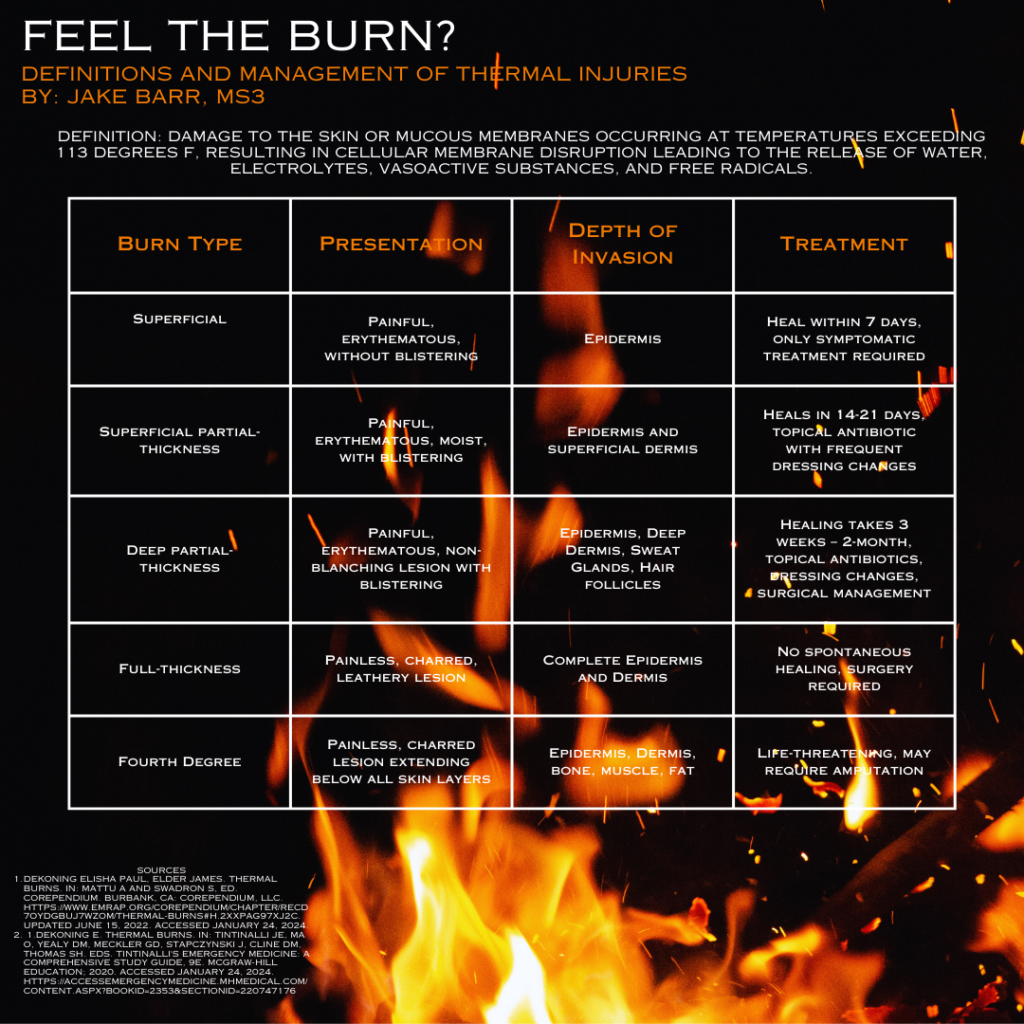
Feel the Burn?

Additional Considerations

· Any patients with concerns of airway burns should be considered for intubation.

· Calculate total surface area burned.

Rules of Nine or Lund-Browder

· After initial fluid resuscitation, use Parkland formula to calculate fluid resuscitation over first 24 hours.

· Provide appropriate analgesia.

· Address tetanus vaccination.

· Wound care is highly dependent on local burn center protocols and guidelines.

Friday Board Review

Board Review by Dr. Edward Guo (Edited by Dr. Parikh)

A 66 year old male with a past medical history of type 2 diabetes presents to the emergency department for head injury. He was the restrained driver when he swerved his car to avoid a child that ran into the street to retrieve a ball. He was driving approximately 15 mph in his neighborhood and swerved onto grass. He hit his head on the driver side window and complains of a headache. He was able to ambulate out of the vehicle and denies loss of consciousness, vomiting, or use of blood thinners. Vital signs are normal. On exam, he is neurologically intact and has ecchymosis to the left forehead. Which of the following is appropriate justification to obtain head imaging according to the Canadian CT Head Rule?

A: Dangerous mechanism

B: Headache

C: Patient age 

D: Rule does not apply to this patient

Answer: Rule does not apply to this patient

The Canadian CT Head Rule is a widely used clinical decision tool that emergency physicians frequently utilize to screen for significant head injuries. In the external validation trial, it was found to be 100% sensitive in detecting both clinically important brain injuries and injuries that required neurosurgical intervention. This patient does not meet the inclusion criteria for its use. If he did meet the inclusion criteria, his age would be an appropriate justification of head imaging as a high risk criteria. Headache is not part of the decision tool. His injury also does not meet the medium risk dangerous mechanism. 

MDCalc states, “Patients with minimal head injury (i.e., no history of loss of consciousness, amnesia, and confusion) generally do not need a CT scan. For example, patients over 65 years old may not need a CT scan just based on their age if they do not have the history mentioned above. When a patient fails the CCHR, use clinical judgment on whether a CT scan is necessary.”

Canadian CT Head Rule
Inclusion CriteriaExclusion Criteria
Head injury with GCS 13-15 and at least one of the following:Age < 16 years
    Loss of consciousnessUse of blood thinners
    Amnesia to head injury eventSeizure after injury
    Witnessed disorientation

References:

Stiell IG, Wells GA, Vandemheen K, et al. The Canadian CT Head Rule for patients with minor head injury. Lancet. 2001;357(9266):1391-1396. doi:10.1016/s0140-6736(00)04561-x

https://www.mdcalc.com/calc/608/canadian-ct-head-injury-trauma-rule