Wednesday Image Review

What’s the Diagnosis? By Dr. Katie Selman

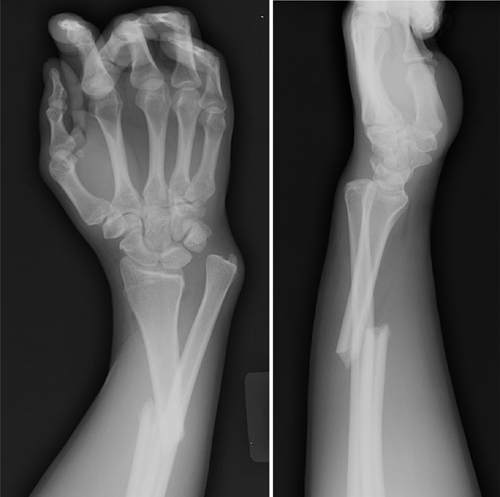
A 63 year old female is brought in by EMS after being found down. She has multiple ecchymoses on her chest and bilateral flanks. GCS is 6. After intubation, she is taken for CT head/cervical spine and a CT chest/abdomen/pelvis with contrast. Upon return from CT, x-rays are done (shown below) to further evaluate bruising and a laceration to her L elbow. What’s the diagnosis?

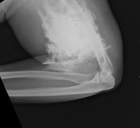
Answer: Contrast extravasation

  • Predisposing factors for contrast extravasation
    • Small IV gauge (22G or less)
    • More distal access (hand)
    • Rapid injection of contrast
  • Incidence: up to 1% of patient receiving IV contrast through peripheral IV
  • Most common symptoms: local pain, swelling
  • Complications occur in < 1 %  (more common with large volume and in patients with atherosclerosis, venous insufficiency, or impaired lymphatic drainage)
    • Compartment syndrome
    • Tissue necrosis
  • Close monitoring required following extravasation
    • Compartment checks, vascular checks, and monitoring of overlying skin
    • Surgery consult for any signs of compartment syndrome or tissue injury
    • Elevate limb, warm compresses may be used
    • Patients rarely require more than conservative supportive treatment

References:

Sbitany, H., Koltz, P. F., Mays, C., Girotto, J. A., & Langstein, H. N. (2010). CT contrast extravasation in the upper extremity: Strategies for management. International Journal of Surgery, 8(5), 384-386. doi:10.1016/j.ijsu.2010.06.002

Sonis, J. D., et al (2018). Implications of iodinated contrast media extravasation in the emergency department. The American Journal of Emergency Medicine, 36(2), 294-296. doi:http://dx.doi.org/10.1016/j.ajem.2017.11.012

Tuesday Advanced Cases & Procedure Pearls

Critical Cases – Hypertensive Emergency!

by Dr. Sarah Perelman M.D.

Today’s case from the EM Daily archives involves one of the rare patients where you DO want to acutely treat elevated blood pressure with intravenous agents….

HPI

  • 48 year old male with PMH HTN presents with blurry vision for 2.5 hours 
  • Patient was using the computer tonight, could not see where the icons were on his desktop, could still see light/colors.
  • He has no pain in his eyes
  • Also reports dyspnea on exertion for 2 days. No headache, no chest pain, no abdominal pain
  • He has not had his anti-hypertensives (he reports he is on 5 different medications) for about 1.5 weeks

Physical Exam

T 98.3 BP 290/120, HR 118, RR 18, SpO2 99%

  • Patient is awake, alert, conversant, appears well and in no distress
  • Neuro: Visual acuity 20/200 OS, OD, OU Normal visual fields Normal pupillary exam Normal extraocular movements Otherwise normal cranial nerve exam Normal strenght in extremities , no pronator drift, normal finger to nose
  • Cardiac: tachycardic, normal S1/S1, no murmurs/rubs/gallops
  • Pulm: clear to auscultation bilaterally
  • Abdomen: soft, nontender, nondistended

Differential Diagnosis 

  • Hypertensive emergency with elevated BP and evidence of end organ damage (decreased visual acuity, evidence of pulmonary edema on bedside US) 
  • Sympathomimetic toxicity (hypertension, tachycardia), though patient reports no ingestions of medications or drugs
  • Thyrotoxicosis 
  • CVA given visual changes, however with no focal visual deficits (no visual field cut, decreased acuity is symmetric bilaterally) 

Initial ED Management 

  • Arterial line place – IV nicardipine started, with goal SBP 210s (25% reduction in the first hour)
  • Bedside lung US performed which demonstrates numerous B lines consistent with evolving pulmonary edema

Labs/Imaging –

  • Hb 6.1, PLT 142, WBC 5.92 – Na 147, K 3.7 – Cr 15.03 (last level in chart 3.95 7 years ago) – HS troponin 223 – pro-BNP 26,930
  • CT Head with 3 small, distinct areas of intraparenchymal hemorrhage

Further Management 

  • Repeat neurologic exam performed and is unchanged
  • Neurosurgery consulted, recommend BP goal under SBP 160
  • Repeat CTH in 4 hours: unchanged 
  • Patient admitted to ICU for IV nicardipine, continuous BP monitoring, and q1 hour neuro checks

Pearls 

  • Hypertensive emergency is acute SBP over 180 with evidence of organ dysfunction
  • Not every patient with SBP over 180 requires emergency BP control
  • In this patient: decreased visual acuity, pulmonary edema, elevated troponin and proBNP, renal failure, and intraparenchymal hemorrhage = hypertensive emergency
  • In managing hypertensive emergency, SBP should not be lowered by more than 25% in the first hour to prevent causing hypoperfusion and cerebral ischemia 
  • Continuous BP monitoring via arterial line is important to carefully titrate medications
  • Nicardepene is an easy to titrate CCB which may be the ideal agent for the treatment of hypertensive emergency
  • Indications for emergent dialysis (AEIOU – acidosis, electrolytes, intoxication, overload, uremia): critical metabolic acidosis, refractory or rapidly increasing hyperkalemia, life threatening intoxication with substance that is able to be removed with HD, volume overload, complications of uremia (pericarditis, neuropathy, encephalopathy)
Monday Back to Basics & Pharmacology

Transvenous Pacing

The last two weeks, Dr. Cash taught us about bradycardia and transcutaneous pacing. This week, we dug back years into the archives and pulled out Dr. Pelletier-Bui’s famous post on transvenous pacing.

A timeless classic! Just be sure to know what supplies are available in your department, where to find them, and alternate options if your supplies differ.

References:

Bessman, E. (2019). Emergency cardiac pacing. In Roberts et al (Eds.), Roberts and Hedges’ clinical procedures in emergency medicine and acute care (pp. 288-308).  Elsevier, Inc.

Bohanske. (2013, November 4). Transvenous pacemaker placement – Part I: The walkthrough.  Taming the Sru.  https://www.tamingthesru.com/blog/procedural-education/transvenous-pacemaker-placement-part-1-the-walkthrough 

Mason, J. [EM:RAP Productions] (2018, October 11).  Placing a transvenous pacemaker [Video]. YouTube. https://www.youtube.com/watch?time_continue=24&v=00-T8PcbStE&feature=emb_title

Friday Board Review

Board Review by Dr. Edward Guo (Edited by Dr. Parikh)

A 30 year old male presents with a displaced right ankle bimalleolar fracture. He is undergoing procedural sedation in the emergency department using midazolam and fentanyl for fracture-dislocation reduction. During the procedure, he becomes apneic and hypoxic. The hypoxia improves with bag valve ventilation, but he becomes progressively more difficult to ventilate. There is absence of chest rise despite increasing positive pressure. What is the likely cause of this patient’s presentation?

A: Laryngospasm

B: Musculoskeletal stiffness

B: Opioid induced hypoventilation

C: Pneumothorax

Answer: Musculoskeletal stiffness

This patient is likely experiencing Rigid Chest Syndrome, a rare but potentially fatal side effect of synthetic opioids causing skeletal muscle rigidity. The exact mechanism is unknown but is related to the dose and administration. It is commonly seen at high doses (> 3 mcg/kg of fentanyl) and with rapid IV push but has been reported with low doses as well. Treatment includes use of propofol for muscle relaxation or naloxone for reversal of opioid agonism. Neuromuscular paralysis and intubation may be required in refractory cases.

Laryngospasm is a known adverse reaction of ketamine administration which usually responds to first-line maneuvers such as jaw thrust or bag valve ventilation. Hypoventilation is a common side effect of opioids but should not cause chest wall rigidity. While uncommon, a pneumothorax may be caused by excessive positive pressure, but at least unilateral chest rise should be visualized with ventilation.

References:

Myers JG, Sutherland J. Procedural Sedation and Analgesia in Adults. In: Tintinalli JE, Ma O, Yealy DM, Meckler GD, Stapczynski J, Cline DM, Thomas SH. eds. Tintinalli’s Emergency Medicine: A Comprehensive Study Guide, 9e. McGraw-Hill Education; 2020.

Çoruh  B, Tonelli  MR, Park  DR: Fentanyl-induced chest wall rigidity case report. Chest 143: 1145, 2013. [PubMed: 23546488.

Patel, Nishika. “Wooden Chest Syndrome.” CriticalCareNow, 5 Aug. 2021, criticalcarenow.com/wooden-chest-syndrome/. Accessed 22 Mar. 2024.

Wednesday Image Review

What’s the Diagnosis? By Dr. Daniel Petrosky

50-year-old with a history of T2DM, hypertension, hyperlipidemia, substance use, and asthma who presents with 4 weeks of progressive left index finger pain. The patient had some swelling from the fall, but now has had one week of swelling worsening, pain, redness. Physical exam reveals digit red, swollen, fusiform, very tender, and unable to flex or extend DIP and PIP. A POCUS is obtained and shown below. What’s the diagnosis?

Answer: Necrotizing Fasciitis

POCUS is very quick to evaluate for many of differentials but would be painful to press directly on the digit with gel. There is an excellent alternative here: waterbath technique.

Water is an excellent medium for ultrasound. In this instance, we submerged the patient’s hand in water, and we are able to submerge the probe and hover above the affect area without concern for the amount of gel we displace in assessing the area or the pressure we apply to get clear images. Also, the high transmissibility of ultrasound through water allows for excellent visibility of superficial structures of body parts like fingers when direct contouring with gel would be virtually impossible. With this technique, we can look for fractures, abscesses, air, and cobble-stoning quickly, easily and with a high degree of accuracy.

So, in this patient as it turns out, air and cellulitis are visible, likely being a necrotizing infection. POCUS with a waterbath was the quickest way to establish this diagnosis. This is extremely important because even with advances in antibiotics, time to surgical intervention is most important in decreasing morbidity and mortality in these patients.

Resources:

Blaivas M, Lyon M, Brannam L, Duggal S, Sierzenski P. Water bath evaluation technique for emergency ultrasound of painful superficial structures. Am J Emerg Med. 2004 Nov;22(7):589-93. doi: 10.1016/j.ajem.2004.09.009. PMID: 15666267.

Shelhoss S C, Burgin C M (November 16, 2022) Maximizing Foreign Body Detection by Ultrasound With the Water Bath Technique Coupled With the Focal Zone Advantage: A Technical Report. Cureus 14(11): e31577. doi:10.7759/cureus.31577

Shrimal P, Bhoi S, Sinha TP, Murmu LR, Nayer J, Ekka M, Mishra P, Kumar A, Trikha V, Aggarwal P. Sensitivity and specificity of waterbath ultrasound technique in comparison to the conventional methods in diagnosing extremity fractures. Am J Emerg Med. 2022 Mar;53:118-121. doi: 10.1016/j.ajem.2021.12.067. Epub 2022 Jan 2. PMID: 35016093.

Bonne SL, Kadri SS. Evaluation and Management of Necrotizing Soft Tissue Infections. Infect Dis Clin North Am. 2017 Sep;31(3):497-511. doi: 10.1016/j.idc.2017.05.011. PMID: 28779832; PMCID: PMC5656282.

Tuesday Advanced Cases & Procedure Pearls

Neonatal Tachycardia by Dr. Edward Guo

Case: A 4 day old female born at 36w1d via C-section presents for tachycardia. Father at bedside states that he placed a heart monitor on the patient and it read over 200 bpm. Patient was seen at urgent care and transferred to ED via ambulance. Baby has otherwise acting normally, eating frequently, making 6+ wet diapers per day. No prior medical history. No family history of abnormal heart rhythms. Vitals include BP 85/53, HR 300, Temp 97.5F, RR 50, SpO2 100%. Exam shows a well appearing, interactive neonate with tachycardia on auscultation. 

Differential diagnosis: Arrhythmia, dehydration, anemia, infection, hyperthyroidism

EKG is obtained and shown below:

EKG interpretation: Supraventricular tachycardia at rate of 300

Case continued: Patient is placed on continuous cardiac monitoring. Vagal maneuvers are attempted including rectal temperature without success in terminating SVT. An IV is placed and adenosine is administered. Repeat EKG following adenosine is shown below. Pediatric cardiology is consulted and recommends administration of digoxin and transfer for further evaluation. Patient is then transferred to a pediatric center in stable condition.

Learning points:

  • A heart rate > 220 bpm in an infant or > 180 bpm in a child with rate out of proportion to clinical status is likely supraventricular tachycardia.2
  • Vagal maneuvers should be attempted initially for patients who are stable.
    • Maneuvers that can be attempted in infants include obtaining a rectal temperature or applying a cold ice pack to the face to activate the mammalian diving reflex.
  • If vagal maneuvers fail, rapid push of IV adenosine at a dose of 0.1 mg/kg is recommended.
  • If the patient is unstable or adenosine fails to terminate the rhythm, perform synchronized cardioversion at a dose of 0.5-1 J/kg.
    • Subsequent synchronized cardioversion attempts should be dosed at 2 J/kg.

References:

Hauda, II WE. Resuscitation of Children. In: Tintinalli JE, Ma O, Yealy DM, Meckler GD, Stapczynski J, Cline DM, Thomas SH. eds. Tintinalli’s Emergency Medicine: A Comprehensive Study Guide, 9e. McGraw-Hill Education; 2020. 

Kleinman  ME, Chameides  L, Schexnayder  SM,  et al: Part 14: pediatric advanced life support. 2010 American Heart Association guidelines for cardiopulmonary resuscitation and emergency cardiovascular care. Circulation 122(18 suppl 3): S876, 2010. [PubMed: 20956230]  

Monday Back to Basics & Pharmacology

Bradycardia, Part 2 with Dr. Allison Cash

  1. Treat possible underlying cause if known 
  1. Atropine
    •  Class 2a treatment
    • Dose 0.5 mg IV every 3-5 minutes, up to total dose 3 mg 
    • Can produce paradoxical worsening bradycardia 
    • Less effective in patients with cardiac transplant 
    • Skip if unstable, move directly to pacing 
  1. Transcutaneous pacing  
    • Class 1a recommendation
    • Place pads in anterior lateral or anterior posterior position 
    • Consider sedation or pain control  
    • Set monitor to “Pacer” 
    • Set rate, usually around 60 bpm 
    • Start current at 0 mA and slowly increase until capture is obtained. Consider different placement of pads if capture not achieved by 130 mA 
    • Confirm capture by feeling pulse or cardiac ultrasound 
    • Transcutaneous pacing is ultimately a bridge to transvenous pacing or permanent pacemaker – discuss with cardiology or transfer to center with these capabilities  
    • Capture (pictured below) shows pacer spike followed by a wide QRS and then ST and/or T wave  

References: 

  1. Judith E. Tintinall, et al. (2020). Tintinalli’s Emergency Medicine : A Comprehensive Study Guide (Ninth Edition). New York: McGraw-Hill. 
  1. Doukky R, Bargout R, Kelly RF, Calvin JE. Using transcutaneous cardiac pacing to best advantage: How to ensure successful capture and avoid complications. J Crit Illn. 2003 May;18(5):219-225. PMID: 30774278; PMCID: PMC6376978. 
  1. Holger J S, Lamon R P, and Minnigan H J et al.: Use of ultrasound to determine ventricular capture in transcutaneous pacing. Am J Emerg Med.  2003; 21: 227 
Friday Board Review

Board Review by Dr. Alex Hilbmann (Edited by Dr. Parikh)

A 66 year old female reports to the emergency department with right arm pain after slipping on ice and trying to catch herself while falling forward. The patient is in incredible pain and has an obvious deformity of the right forearm upon presentation. An x-ray of the right forearm is shown below. What injury does the patient have?

A. Colles Fracture

B. Monteggia Fracture

C. Galeazzi Fracture

D. Smith Fracture

Answer is C. The x-ray shows a distal third radial fracture with disruption of the distal radioulnar joint space, which is a Galeazzi fracture. A Colles Fracture (A) is a distal radius fracture with dorsal displacement whereas a Smith Fracture (D) is a distal radius fracture with volar displacement. Both fractures frequently do not involve significant disruption of the radioulnar joint. A Monteggia fracture (B) is a fracture of the proximal ulna which results in radial head dislocation. For a Galeazzi fracture, the anterior osseous nerve (a branch of the median nerve) is often affected and function should be assessed by asking the patient to perform an “Okay” sign with first and second digit of affected arm.  Orthopedic Surgery consultation is necessary in adults as treatment is an open reduction and internal fixation (ORIF). If open fracture is present Cefazolin should be administered and if the wound is > 10 cm, appears contaminated, or involves seawater/freshwater/farming equipment Gentamicin should also be administered.

Picture from:

https://www.orthobullets.com/trauma/1029/galeazzi-fractures

Resources:

Atesok KI, Jupiter JB, Weiss AP. Galeazzi fracture. J Am Acad Orthop Surg. 2011 Oct;19(10):623-33. doi: 10.5435/00124635-201110000-00006. PMID: 21980027.

Garg R, Mudgal C. Galeazzi Injuries. Hand Clin. 2020 Nov;36(4):455-462. doi: 10.1016/j.hcl.2020.07.006. PMID: 33040957.

Mills, Trevor J.. “Forearm Fractures.” (2013). https://www.semanticscholar.org/paper/Forearm-Fractures-Mills/dead0398468fc50a88349251a8cb8a49b88f838f